


A Novel Method to Generate Colon Cancer Orthotopic Tumors in Mice: Implantation Using the Cecal Pouch Technique
Carlos H. Chan*1,2, Anne-Laure Nouvion2, Luisa Demarte2, ValéRie Breton2, Claire Turbide2, Nicole Beauchemin2, Lorenzo E. Ferri1
1Department of Surgery, McGill University, Montreal, QC, Canada; 2Goodman Cancer Research Centre, McGill University, Montreal, QC, Canada
Background: The ectopic subcutaneous tumor model is the most commonly used pre-clinical model for cancer drug development, but disregards the importance of primary tumor microenvironment. Studies have shown that cancer progression and responses to chemotherapy are different between ectopic and orthotopic tumors. Cecal microinjection of cancer cells is the standard orthotopic colon tumor model, but major concerns have been cited regarding reliability and technical difficulties with this method. We here describe a novel and highly reliable technique of developing orthotopic colon cancer in mice.Methods: Using C57BL6 mice, midline abdominal incisions were made and small pouches were created at the tip of the cecums by invaginating the bowel wall (Fig. 1b). Intact tumor fragments (approximately 2mm x 2mm x 2mm) derived from liver metastases of MC-38 cells (mouse colon cancer cell line) were enveloped within the cecal pouches, which were then closed with 3-0 silk ties (Fig. 1c). Mice were monitored over a 6-week period. At experimental or clinical endpoints, the abdominal cavities were examined for the presence of cecal tumors, gross liver and lymph node metastasis, and carcinomatosis. The size of cecal tumors and number of liver surface metastatic nodules were also determined. Tumors and metastatic nodules were examined by routine histology.Results: The average operating time was approximately 10 minutes per mouse using this novel cecal pouch technique. The 6-week survival was 77% (10/13); 2 died without any tumor within the first 10 days and 1 with cecal tumor at 4 weeks. Of the 10 mice surviving to study completion, 8 harbored tumors within their cecal pouches (Fig. 2) and 2 had generalized carcinomatosis secondary to perforated cecal pouches. Successful generation of orthotopic tumor was seen in 85% (11/13) of mice. Liver metastases were detected in 3 mice without carcinomatosis (Fig. 2a). Using histological analysis, all non-perforated cecal tumors (8/8) involved entire bowel wall (mucosa, submucosa and muscularis propria), but were still enclosed by the outer pouch layer, representing T3 tumors.Conclusion: Our novel cecal pouch approach may represent an efficient method to generate clinically relevant orthotopic cecal tumors for quantitative analysis and pre-clinical studies.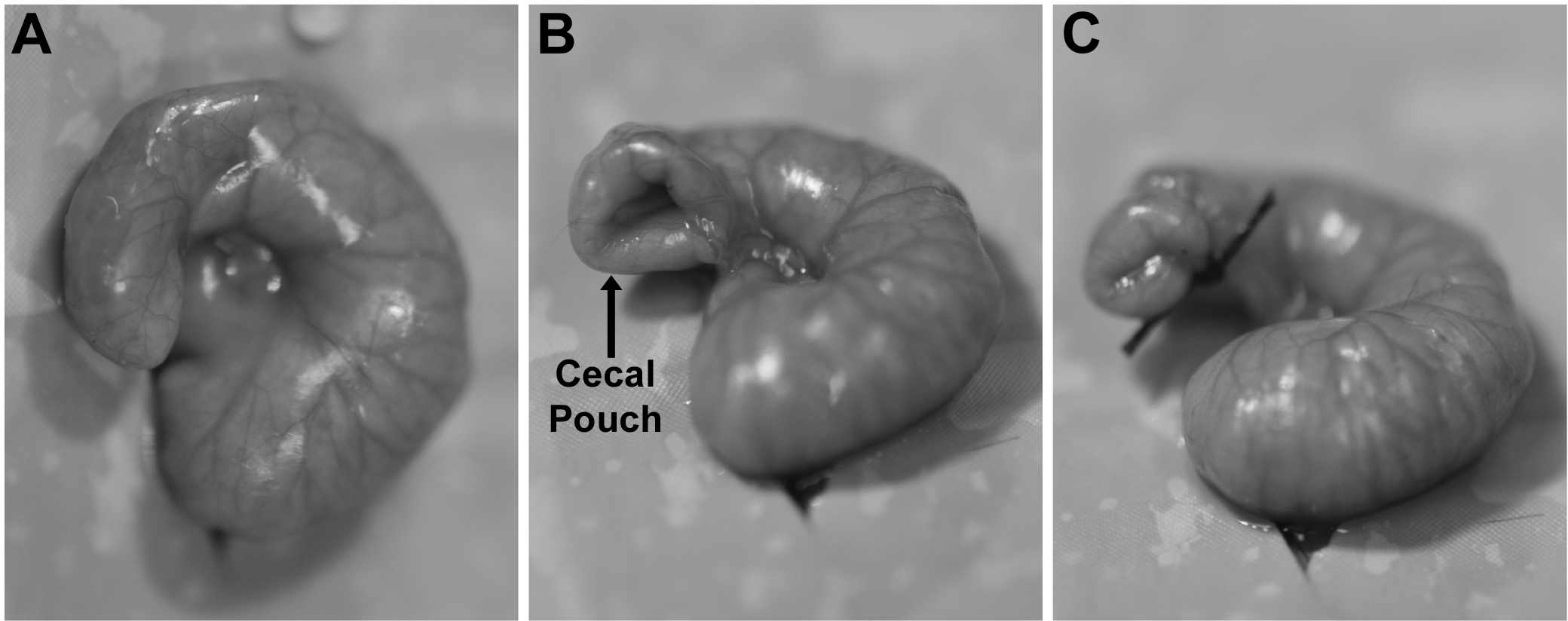
Cecal pouch technique for tumor implantation: A) Exteriorized cecum through midline abdominal incision; B) Creation of cecal pouch by invaginating the bowel wall; C) Closure of cecal pouch by 3-0 silk tie.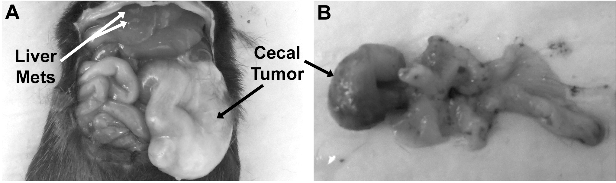
Mouse with a large tumor enclosed in the cecal pouch (black arrow) and gross liver metastases (white arrows) at 6-week post-implantation.
Back to 2011 Program