
EX-VIVO HUMAN MODEL OF THERANOSTIC NANODELIVERY SYSTEM FOR PANCREATIC NEOPLASMS - A FEASIBILITY TRIAL
Konstantinos Chouliaras*, LACEY MCNALLY, Abhilash Samykutty, Libby McWilliams, Ralph D'Agostino, Clancy J. Clark, Perry Shen
Wake Forest Baptist, Winston-Salem, NC
Introduction
Despite the progress made in the diagnosis and treatment of pancreatic neoplasms, there remains considerable ambiguity and false negative results. Multispectral Optoacoustic Tomographic Imaging (MSOT) has been used in-vitro and in animal models targeting pancreatic cancers showing good affinity. In this trial we sought to demonstrate the affinity of the nanoparticles in human pancreas tumor specimens.
Methods
Patients who underwent resection of pancreatic tumors were prospectively enrolled in the trial. 0.5 x 0.5 x 0.5 cm of fresh tumor and normal tissue (cells and stroma) were obtained during a resection with curative intent.
An acidic pH targeted wormhole mesoporous silica nanoparticle, coated with chitosan and targeted with a V7 peptide (V7-CWMSN) was developed to serve as a contrast agent. The acidic pH targeted particle, V7-CWMSN, containing either IR-780 dye or propidium iodide was evaluated in human pancreatic tumor and normal tissue. Tumor uptake of the particle was evaluated using MSOT with secondary confirmation using near infrared fluorescent imaging (NIR).
Results
Four patients were included in this feasibility trial - 2 underwent Whipple resections and one distal pancreatectomy for pancreatic adenocarcinoma and another patient underwent Whipple for isolated breast carcinoma metastases to the pancreas.
The core mesoporous component of the particle was 33.0±1.94 nm and enlarged to 38.87 ±1.5 nm upon the addition of the chitosan gatekeeper. The polydispersity index indicated a monodispersed particle at 0.12 over the course of 100 h indicating a lack of particle aggregation. Ex vivo evaluation of particle uptake in resected patient pancreatic tumors at pH 6.6 resulted in 2.3X increased uptake compared to normal tissue at pH 7.4 (p=0.0023) as observed using 3D MSOT imaging and 1.9 X increased uptake at pH 6.6 compared to pH 7.4 (p=0.0076) using 2D NIR fluorescence imaging.
Conclusions
In this feasibility trial, increased affinity of the mesoporous silica nanoparticle was shown both with MSOT and NIR fluorescence imaging. Further study is needed to confirm the reproducibility of these results.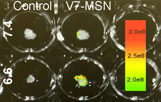
Figure. Near infrared fluorescent imaging showing high affinity of the pH-targeted V7-CWMSN nanoparticle to pancreas tumor compared to normal tissue.
Back to 2019 Posters


