
CLINICOPATHOLOGIC STUDY OF SURGICAL MARGIN FOR LIVER RESECTION USING NEAR-INFRARED FLUORESCENT IMAGING AND FLUORESCENT MICROSCOPE
Takahito Hirai*, Takeshi Aoki, Tomotake Koizumi, Tomokazu Kusano, Akira Fujimori, Yoshihiko Tashiro, Koji Nogaki, Tomoki Hakozaki, Kodai Tomioka, tatsuya yamazaki, Yusuke Wada, Kazuhiro Matsuda, Hideki Shibata, Marie Uchida, Yuta Enami, Masahiko Murakami
Gastroenterological & General Surgery, Showa University, Shinagawa-ku, Japan
Objectives
Near-infrared fluorescent (NIR) imaging has been widely applied across various fields of surgery. Here, we analyzed the fluorescence signals from liver tumor in the transection plan using NIR imaging, considering it to be a reliable indicator for securing surgical margin. This study intended to histologically validate the observation technique using NIR imaging to secure safety surgical margin histologically.
Method
From 2014 to 2017, patients with liver tumors underwent hepatectomies under the guidance of NIR imaging. ICG was administrated (0.5 mg/kg) 3-14 days prior to the surgery for the observation of tumor location and transection plane. Hepatectomies (n = 42; limited liver resection) were evaluated using NIR imaging, and the liver parenchyma was carefully transected so as to not expose the fluorescent signal from the tumors. Histopathological examination for hepatocellular carcinoma (HCC) and metastatic colorectal cancer (CRLM) cases was performed under a fluorescence microscope (BZ-X800: Keyence, Osaka, Japan) equipped with a near-infrared light source.
Result
Surgical specimens from 16 cases of HCC and 26 cases of CRLM were histopathologically evaluated under a fluorescence microscope. The fluorescence patterns of HCC were as follows: total fluorescent type, 5; partial fluorescent type, 1; rim fluorescent type, 7; and combined type, 3 (Partial + Rim,1; Total + Rim; 2).
In the cases of HCC, the fluorescence range was set at ≥5 mm from the tumor margin in 4 cases, <5 mm in 6 cases, and fluorescent only within the tumor in 6 cases. Conversely, the fluorescence pattern of CRLM was Rim fluorescent type (i.e., a fluorescence signal only surrounding the tumor) in all cases. The fluorescent range for CRLM was >5 mm in 8 cases and <5 mm in 18 cases. In the cases of both HCC and CRLM, under fluorescence microscope, no malignant finding was determined histopathologically in the area with fluorescence signal surrounding the tumor.
Conclusion
Histopathological findings suggested that the region with fluorescent signal surrounding the liver tumor did not contain malignant cells. Unless a fluorescence signal was exposed in the transection plane of the liver, it was important to remove all fluorescent portions in the remnant liver for securing curability. This study histopathologically validated that NIR imaging is a reliable intraoperative navigation tool for identifying surgical margins for liver surgery.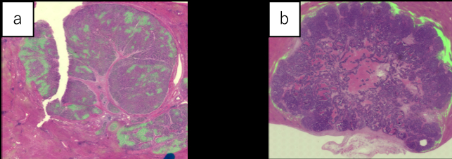
a:HCC b:CRLM
Back to 2019 Abstracts


