|


|
Back to Annual Meeting Posters Improving the Detection of Mitoses in Pancreatic Neuroendocrine Tumors Using Phosphohistone H3 Stephanie L. Goff*1, Matteo Ligorio3, Jennifer a. Wargo1, Zachary Cooper1, Dennie T. Frederick1, Francesco Sabbatino3, Vikram Deshpande2, Cristina R. Ferrone3 1Surgical Oncology, Massachusetts General Hospital, Boston, MA; 2Pathology, Massachusetts General Hospital, Boston, MA; 3General Surgery, Massachusetts General Hospital, Boston, MA
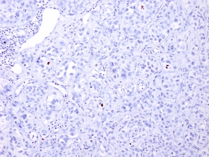
Pancreatic neuroendocrine tumors (PNET) are being diagnosed with greater frequency and have a widely variable natural history. Recent advances in staging for PNET incorporate the mitotic rate for tumors identified on hematoxylin and eosin stains (H&E), dividing lesions into low grade, intermediate grade, and high grade based on mitotic rate. This staging system can help with prognostic information, and may also guide adjuvant treatment, but is reliant on an accurate assessment of mitotic count. By H&E, mitotic figures can be difficult to identify and require an experienced histopathologist for accurate enumeration. Proliferative index, as measured by Ki-67, is also used in PNET but may over-represent actual mitotic figures since this also captures the G1 phase. More recently, staining with phosphohistone H3 (PHH3) has been described for use in identifying mitotic figures in malignancies such as melanoma and glioblastoma. PHH3 is exclusively expressed in the mitotic phase of the cell cycle thus yields a much more accurate representation of mitotic rate in tumors. Our hypothesis was that an anti-PHH3 antibody can more accurately identify mitotic rates in pancreatic neuroendocrine tumors (PNET) than traditional H&E staining. This will result in more accurate staging and will improve patient management. Back to Annual Meeting Posters
|
|||||||
© 2026 Society for Surgery of the Alimentary Tract. All Rights Reserved. Read the Privacy Policy.