Back to 2025 Posters
COLON CANCER OR SOMETHING ELSE? UNMASKING AN APPENDICEAL ADENOCARCINOMA
Udit Nangia
*, Peter Alamir, David N. Linz, Dany Raad
University Hospital - Parma Medical Center, Parma, OH
Background:Primary appendiceal cancers are rare, with an incidence of approximately 1.2 cases per 100,000 population. In contrast, primary colon cancer is far more common, with an incidence of 36 cases per 100,000 population in the United States. Appendiceal cancers often remain asymptomatic until they progress beyond the appendix, making early diagnosis challenging. We present a case of an elderly man initially thought to have colon adenocarcinoma, later identified as a primary appendiceal adenocarcinoma, underscoring the importance of considering rare malignancies in atypical presentations.
Case Presentation:A 78-year-old man underwent a screening colonoscopy, which revealed a 25 mm polyp at the appendiceal orifice. Attempts at endoscopic resection were incomplete due to excessive bleeding and technical difficulty in lifting the lesion. A 9 mm pedunculated polyp in the sigmoid colon was also identified and successfully resected (Image 1).
Pathology of the cecal lesion showed tubulovillous adenoma with high-grade dysplasia, while the sigmoid polyp biopsy confirmed invasive, moderately differentiated adenocarcinoma. A repeat colonoscopy with additional biopsies and tattooing of the sigmoid lesion yielded similar findings. A laparoscopic colectomy was planned with dual resections for presumed synchronous lesions.
Intraoperatively, the tumor was found to originate from the appendix with local invasion into the sigmoid colon (Image 2). This discovery necessitated an en bloc resection of the affected regions. The final surgical pathology confirmed the diagnosis of primary appendiceal mucinous carcinoma with secondary invasion of the sigmoid colon. The patient recovered well and was referred for adjuvant chemotherapy.
Discussion:Adenocarcinoma is the most common primary appendiceal cancer but accounts for less than 0.5% of gastrointestinal malignancies. Subtypes include mucinous, colonic-type, and signet-ring adenocarcinomas, with mucinous variants differing histologically and physiologically from colonic adenocarcinomas.
In this case, initial findings pointed to synchronous colonic lesions, but surgical exploration revealed a locally invasive primary appendiceal tumor. This highlights a frequent challenge in appendiceal cancer care: preoperative misdiagnosis. While surgical resection remains the cornerstone of treatment, no consensus exists regarding the optimal extent of resection. Adjuvant chemotherapy is often employed for advanced cases.
This case underscores the importance of maintaining a broad differential diagnosis when managing gastrointestinal malignancies. Early recognition of rare entities such as appendiceal cancer may rely on advances in endoscopic techniques or the development of specific biomarkers. Furthermore, operative exploration plays a crucial role in defining the true extent of disease and guiding appropriate management strategies.
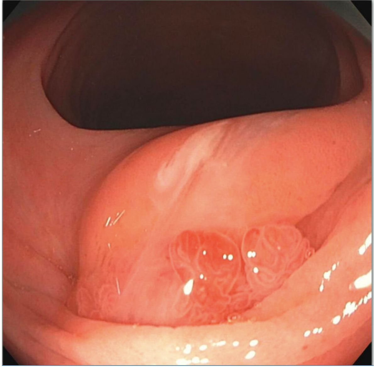
Image 1. Endoscopic image of a 9 mm pedunculated polyp in the sigmoid colon.
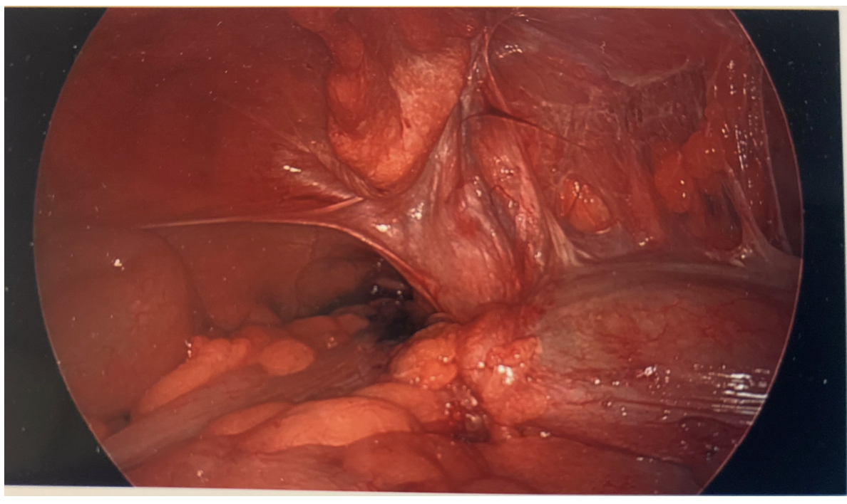
Image 2. Laparoscopic image of appendiceal tumor with local invasion to sigmoid colon.
Back to 2025 Posters


