Introduction
The swine is valuable model for preclinical research and surgical technique training. From the induction of Diabetes through total pancreatectomy, these animals can be used in many research studies, such as islet transplantation. In view of lack of elucidation in the literature, the purpose of this work is to describe anatomical aspects, surgical technique of total pancreatectomy, intra- and postoperative complications and autopsy results.
Material and Method
Five male pigs, hybrids (Large White/Landrace), 20-35 kg, submitted to total pancreatectomy with duodenum, bile duct and spleen preservation. Postoperatively, daily clinical assessment and capillary blood glucose collection were performed. At the end of the 30-day limit or when serious clinical complications occurred, euthanasia and autopsy were performed.
Results
The average duration of surgery was 128 minutes, without intraoperative deaths or anesthesia induction failures. Median survival was 6.6 days. Postoperative complications were weight loss (3), emesis (2), constipation (2), abdominal distension (2), diarrhea (1) and loss of appetite (1). All animals were euthanized due to serious complications. Two animals presented surgical complications (duodenal necrosis with gastroparesis and internal hernia with intestinal necrosis). The other three animals presented serious clinical complications related to exocrine pancreatic insufficiency due to deficiency of pancreatic enzymes. Glycemic values above 200 mg/dL were found on the first postoperative day and above 300 mg/dL on the seventh day in all pigs.
Discussion and Conclusions
In this experiment, we established a model of total pancreatectomy with preservation of the duodenum and spleen in pigs in which all animals had diabetes, however animals without postoperative complications were euthanized due to serious complications related to exocrine insufficiency of the pancreas.
Key words
Pancreas, anatomy, pancreatectomy, swine, experimental model, diabetes, islet transplantation.

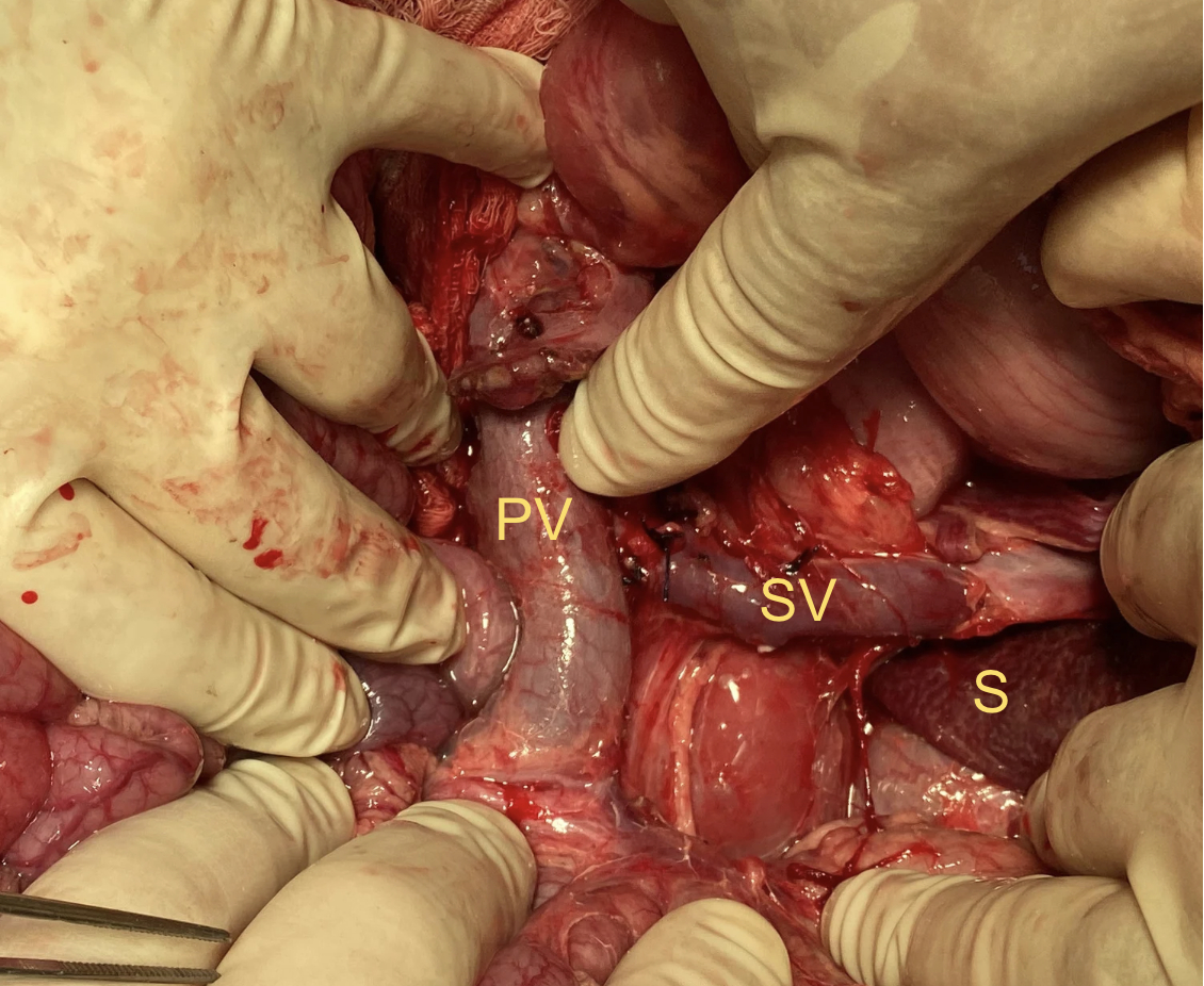
Figure 1: Abdominal cavity after total resection of the pancreas. It is possible to observe adjacent structures such as the portal vein (PV), splenic vein (SV) and spleen (S).

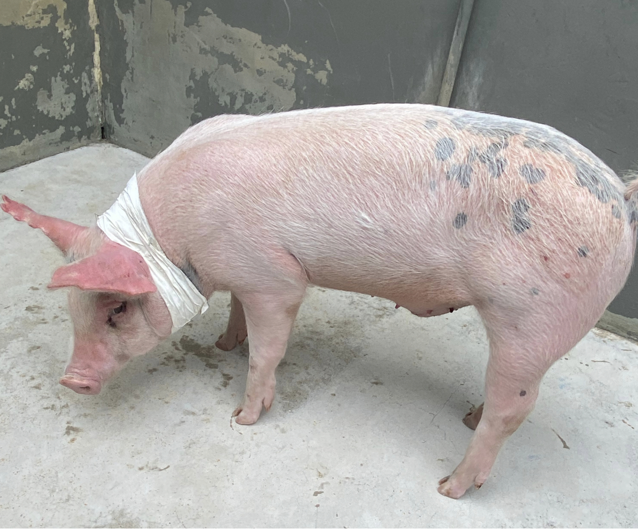
Figure 2: Postoperative period of the third swine. The animal was placed in an individual stall at the bioterium of Faculty of Medicine of the University of São Paulo. The access was maintained in the jugular vein for administration of antibiotic therapy and hydration and blood collection.