
De novo neoplasms are a major complication of solid organ transplant. Describing tumor proliferation in transplant patients is a crucial step in understanding the pathophysiology of tumors in these patients and therefore, to better optimize immunosuppression and screening guidelines. In this case report, we present a granular cell tumor found in a patient six years after liver transplant. The patient, a 72-year-old male, underwent an orthotopic liver transplant in 2017 due to liver failure secondary to hepatocellular carcinoma and hepatitis C, and was placed on a standard immunosuppression regimen of tacrolimus. In August 2023, the patient presented to the emergency department with nausea and vomiting and was found to have a mesenteric mass abutting the appendix on CT scan, which had not been visualized in prior scans before and after transplant. The patient additionally underwent an MRE and PET CT which, of note, was positive. The patient was then referred for surgery where the entire tumor was resected in an open right hemicolectomy in November 2023. Pathology returned indicating that the neoplasm is composed of infiltrating round, polygonal to slightly spindled cells with eosinophilic cytoplasm, fibrosis, and chronic inflammatory cells (Fig 1a). The tumor cells are positive for S100 (Fig 1b) and CD68 (Fig 1c). The morphologic features and immunostaining patterns are most compatible with benign granular cell tumor involving the appendiceal wall. Granular cell tumors are rare tumors that have not been described in transplant patients. They can be benign or malignant and are most commonly found in the skin or soft tissue, with a small percentage arising in the GI tract. In presenting this case, we hope to highlight the unique features of this tumor and bring awareness to a type of GI mass seen in the setting of a prior liver transplantation.
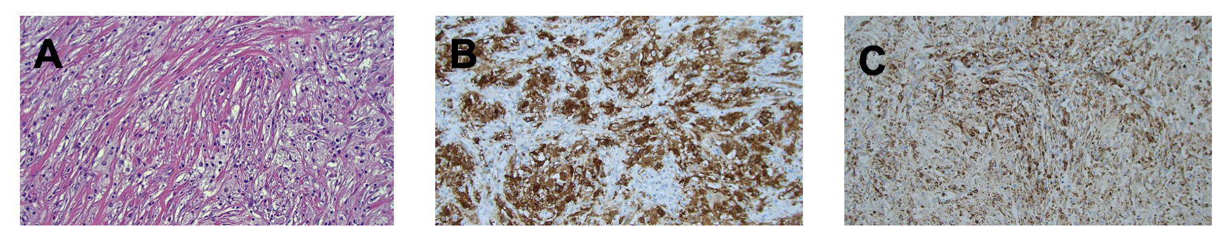
Figure 1. Light microscopy of tumor sample preserved in formalin and stained with H&E (a), s100 (b), and CD68 (c)

