Back to 2024 Abstracts
TUMOROIDS WITH DIFFERENT GENETIC BACKGROUNDS HAVE DIFFERENTIAL RESPONSES TO BILE ACIDS ENRICHED BY A HIGH FAT DIET
Claire E. Wild
*1,2, Zitong Lin
2, Meejeon Roh
2, Colin W. Steele
3, Benjamin D. Shogan
21University of Chicago Pritzker School of Medicine, Chicago, IL; 2The University of Chicago Medicine, Chicago, IL; 3University of Glasgow, Glasgow, United Kingdom
Introduction: A high fat diet (HFD) is a leading environmental risk factor for the development of colorectal cancer (CRC). One of the mechanisms by which a HFD is thought to promote CRC is by enrichment of carcinogenic bile acids. If the carcinogenic activity of bile acids is dependent upon the genetic background of the tumor is largely unknown. The adenoma-carcinoma sequence and the serrated-carcinoma sequence are the two primary mutational sequences that result in CRC. In this study, we sought to determine if HFD-enriched bile acids had a different carcinogenic effect on adenoma-carcinoma tumoroids compared to serrated carcinoma tumoroids.
Methods: C57BL/6 mice were fed either a standard diet (6.2% fat, 3.5% crude fiber) or a HFD (60% fat, 0% crude fiber) for 6 weeks. Excreted stool was collected and immediately stored at -80
oC. Bile acid metabolomics of 40 primary and secondary acids was performed. Bile acids were then tested for their effect on two different tumoroid cell lines: AKPT (
Apcfl/fl;KrasG12D/+;Tp53fl/fl;Tgfbr1fl/f) mimicking the adenoma-carcinoma pathway or KPN (
KrasG12D/+;Tp53-/-;Rosa26N1icd/+) mimicking the serrated-carcinoma pathway. Bile acids that were most enriched by a HFD were co-incubated with either AKPT or KPN tumoroids, and proliferation and volume of tumoroids were assessed using a cell viability kit and by staining with proliferative marker Ki-67. Tumoroid volume was directly measured by tracing individual tumoroids over a 3-day growth period.
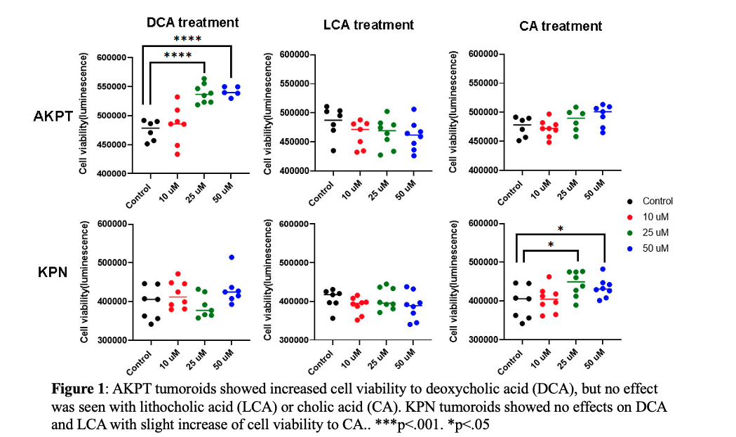
Results: A HFD had a significant influence on the concentration of many bile acids metabolites. The bile acids that were most enriched by a HFD were deoxycholic acid (4811 µg/mL vs 298 µg/mL), lithocholic acid (360 µg/mL vs 24 µg/mL), and cholic acid (87 µg/mL vs 4 µg/mL). These 3 bile acids were then chosen for their effect on AKPT and KPN tumoroids.
We demonstrated that deoxycholic acid (DCA) significantly increased cell viability (mean luminescence ±SD 5.4x10
5 ±7.5x10
3 vs 4.7x10
5 ± 1.3x10
4; p < 0.01) and cell volume (1,394cm
3 ± 529 vs. 805cm
3 ±175; p < 0.01) of AKPT tumoroids but had no significant influence on KPN tumoroids (
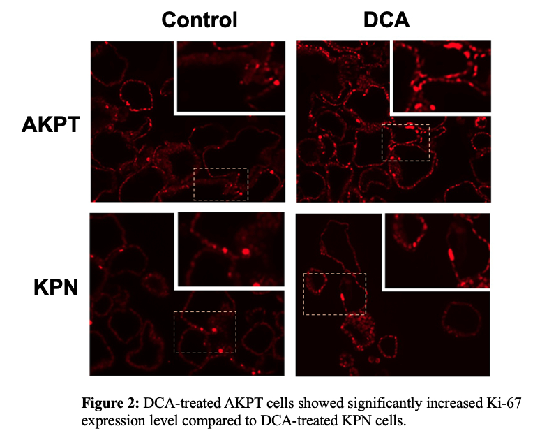
Fig.1). Ki-67 staining in DCA-treated AKPT tumoroids was significantly increased compared to DCA-treated KPN tumoroids (
Fig2). Alternatively, cholic acid promoted cell viability of KPN tumoroids (4.3x10
5 ±1.7x10
4 vs 3.9x10
5 ±3.1x10
4; p<0.05) but had no influence on cell volume or Ki-67 staining in both tumoroids. Lithocholic acid had no effect in any tested assay in either AKPT nor KPN tumoroids.
Conclusion: Bile acids enriched by a HFD had a tumorigenic effect that was dependent upon the genetic background of the tumor. Further research is needed to determine why certain mutations respond to specific bile acids.
Back to 2024 Abstracts


