
PLATELET INDICES AND RED CELL DISTRIBUTION WIDTH IN THE DIAGNOSIS OF ACUTE APPENDICITIS
Gurushankari Balakrishnan*, Premkumar Ramasubramani, Sathasivam Sureshkumar, Noyal M. Joseph, Thulasingam Mahalakshmy, Debdatta Basu, Vikram Kate
Surgery, Jawaharlal Institute of Postgraduate Medical Education and Research, Puducherry, Puducherry, India
Introduction: The rate of negative appendicectomy (10% to 30%) has been stable over the past few decades due to diagnostic dilemma. Studies have focused on determining a simple, cheap, and feasible investigation for an accurate diagnosis based on platelet indices and red cell distribution width (RDW), however their role in the diagnosis is not established. Hence, this study was carried out to determine the role of platelet indices and RDW in the diagnosis of acute appendicitis.
Methods: In this prospective study patients of age >18 years with a clinical and radiological diagnosis of acute appendicitis were included. The patients of appendicitis were grouped into four categories histopathologically normal appendix, uncomplicated and complicated acute appendicitis, and conservative group where appendicitis was diagnosed radiologically. Hematological parameters such as hemoglobin, total leucocyte count (TLC), differential leucocyte count (DLC-N), absolute neutrophil count (ANC), platelet count, mean platelet volume (MPV), platelet distribution width (PDW), RDW and were correlated between the groups.
Results: A total of 237 patients were included in this study. Among the hematological parameters TLC (p=0.0003), DLC-N (p<0.001), and ANC (p=0.0002) were significantly higher in positive appendicectomy group. However, MPV, PDW and RDW were similar between the positive and negative appendicectomy group. Univariate analysis on significant parameters revealed gender (p=0.004), TLC (p=0.001); DLC-N (p=0.001); DLC-L (p=0.001); ANC (p=0.001) were associated with acute appendicitis. (Table 1) Multivariate analysis showed no significance except gender as all parameters were components of TLC. The overall accuracy as determined by receiver operating characteristic (ROC) curve showed that area under the curve (AUC) was good for ANC (0.73; CI: 0.62-0.83), TLC (0.71; CI: 0.61-0.81) and for DLC-N (0.70; CI: 0.58-0.83). (Table 2)
Conclusion: The study showed that MPV, PDW and RDW were not indicative of acute appendicitis, however, higher TLC, DLC-N, and ANC is diagnostic of acute appendicitis as a combined tool.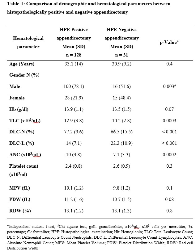
Table-1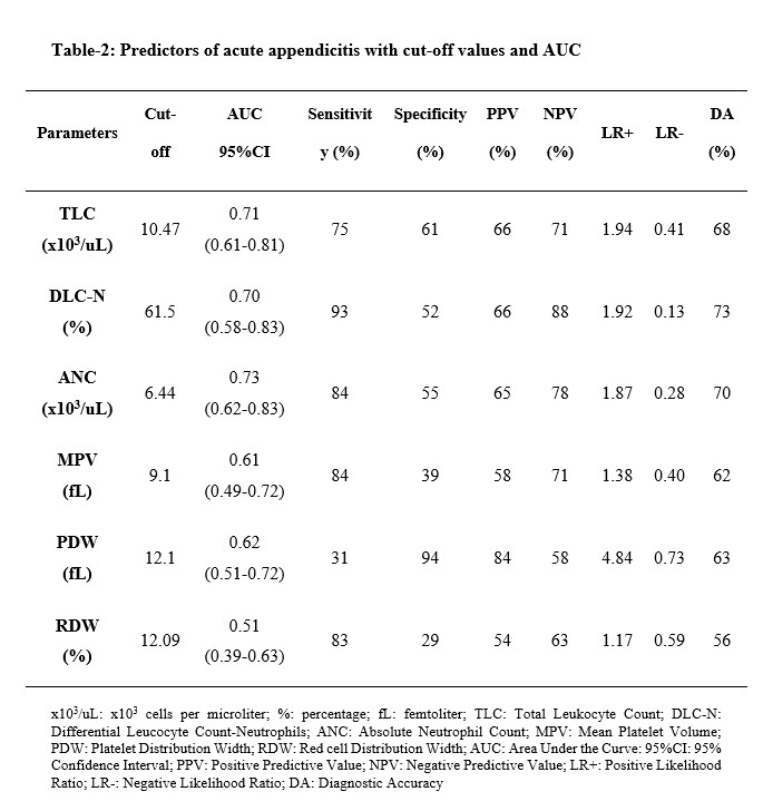
Table-2
Back to 2022 Posters