RIGHT HEPATECTOMY FOR GIANT LIVER HEMANGIOMA GUIDED WITH INTRAOPERATIVE ULTRASOUND AND INDOCYANINE GREEN
Layla Lissette Monroy Velasco1, José David C. Zamora1, Gloria C. Salgado1, Maria Fernanda Arboleda1, Eduardo E. Montalvo-Javé2, Yukiyoshi K. Fujikami3, Alejandro Rossano García*1
1Surgery, Hospital Angeles Pedregal, Mexico City, Mexico City, Mexico; 2Medica Sur, Ciudad de Mexico, Distrito Federal, Mexico; 3CT Scanner Lomas Altas, Mexico, Mexico
Introduction
Hepatic hemangiomas are the most common benign liver tumor, with prevalence of 0.4- 7.3 and an incidence of 0.4-20% in the general population. It is typically observe in females than male 5:1 at age 50 years.
The etiology is unclear, hypothesis suggest they result from abnormal angiogenesis. Hormones such as estrogens play an important role in growth, since they appear more frequently during pregnancy and hormonal therapy.
Most are usually asymptomatic, often diagnosed incidentally, symptomatic ones are associated with larger lesions, according to their dimension they can be small or giant (>5cm).
Indications for surgery include progressive abdominal symptoms, high grade of rupture, rapidly enlarging lesions, Kasabach- Merrit syndrome or suspect of malignancy. Surgical management includes enucleation, hepatic artery ligation, liver resection and liver transplantation.
Case presentation
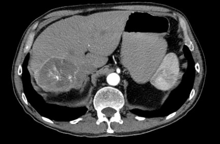
A 73-year-old man with COPD and two previous small hemangiomas. Presents oppressive pain in the right upper quadrant in July 2021. His serum tumor markers were in normal limits. Abdominal CT showed nodular images with an enhancement pattern in segment IV and V of 2.5x2 cm and 9x8.2x4.3cm in the right lobe, compatible with cavernous hemangiomas
Due to the size and symptoms, it was decide to perform the right hepatectomy guided with indocyanine green and intraoperative ultrasound
Discussion
We opted for elective resection; with an open approach. Compared with laparoscopic, although feasible, this would exacerbate the preexisting pulmonary restriction.
Using the bipolar sealers combining radiofrequency energy and saline to provide a hemostatic sealing of the soft tissue, consequently controlling bleeding and reducing the surgical time. The patient presented a bleeding of 500cc with minimal blood loss, as it was a highly vascularized tumor; only one globular package was transfuse.
Tissues such as vascular structures present a higher resistance due to their high collagen content; the use of the ultrasonic aspirator provides a greater tactile response when in contact with critical structures, allowing its maximum selectivity during resection.
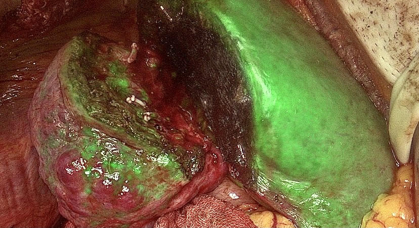
We avoided injury to critical structures and blood vessels in a hemangioma with multiple tributaries, while the use of indocyanine green allowed us to guide adequate perfusion of the remaining liver.
Conclusions
Giant hemangiomas are susceptible to embolization or resection, however, due to their size, it was decide in a surgical event to perform hepatectomy with a lower risk of rupture and multiple interventions.
Open hepatectomy was carried out successfully, free of pulmonary complications and bleeding. On its follow up, he is currently undergoing pulmonary rehabilitation, with a tendency towards normalization of his liver function tests and with a good quality of life, Barthel score 20 points and Katz index grade A.
Indocyanine green with perfusion of complete liver remnant and partially devascularized liver hemangioma
Abdominal CT showing nodular images with an enhancement pattern in the arterial phase, characteristic of hepatic hemangiomas
Back to 2022 Posters
