
INNATE IMMUNITY IN COLORECTAL LIVER METASTASES: MACROPHAGE MORPHOLOGY AND DISTRIBUTION ARE STRONG PREDICTORS OF PROGNOSIS. AN EXTERNAL VALIDATION.
Guido Costa*1,2, Matteo Donadon1,2, Faizan Nasir2, Cristiana Soldani1, Barbara Franceschini1, Michela Anna Polidoro1, Carlo Sposito3,4, Matteo Virdis3, Di Tommaso Luca1,2, Andrea Vingiani3,4, Vincenzo Mazzaferro3,4, Guido Torzilli1,2
1IRCCS Humanitas Research Hospital, Rozzano, Lombardia, Italy; 2Humanitas University, Milan, Italy; 3Fondazione IRCCS Istituto Nazionale Tumori di Milano, Milan, Italy; 4Universita degli Studi di Milano, Milano, Lombardia, Italy
Introduction: Tumor-associated macrophages (TAMs) are key components of tumoral microenvironment and have been shown to impact prognosis in different cancer subtypes. Previously reported data from Humanitas University group showed that TAMs morphology correlates with prognosis in colorectal liver metastases (CLMs), with smaller TAMs (S-TAMs) conferring a more favorable prognosis than larger ones (L-TAMs). This study aims to externally validate TAMs morphology as a prognostic factor in resected CLMs. Moreover, we aim to evaluate the distribution and density of these two populations within the invasive margin of the tumor.
Methods: 86 formalin-fixed and paraffin-embedded post-resection samples of both CLMs and peritumoral tissue were used. Two-?m sections slides were obtained, stained with anti-CD163 and digitalized. With a digital pathology tool, area and perimeter from 21 macrophages in each slide (7 cells from three non-contiguous, non-overlapping different invasive margin zones, selected by a pathologist blinded to the study) were recorded (figure, A).
Results: Average macrophage perimeter was 71.5 ± 14.1 ?m whilst average macrophage area was 217.7 ± 67.8 ?m2. At univariate analysis, TAMs area demonstrated a statistically significant association with DFS (p=0.0006). Optimal area cut-off value was obtained, showing a sensitivity and specificity of 92% and 56%, respectively (Figure, B). S-TAMs and L-TAMs were associated with a 3-year DFS rates of 60% and 8.5%, respectively (P<0.001). Multivariate Cox regression analysis confirmed the strong predictive role of TAMs' area for DFS (HR 6.12, p=0.004), along with the number of resected CLMs (HR 1.12, p=0.006) (Table).
Moreover, in a subset of patients (n=12) characterized by un-favorable (n=6, recurrence within 3 months) or favorable (n=6, no recurrence after 48 months) prognosis, TAMs showed a different distribution: L-TAMs that were more abundant and closer to the tumor invasive margin in patients that encountered early recurrence, and foci of L-TAMs were significantly more extended in patients showing a less favorable prognosis. (Figure, C).
Conclusions: This external validation study confirms that morphometric characterization of TAMs can serve as a simple readout of their diversity and allows to reliably stratify patient outcomes and predict recurrence. Future perspectives could include involve an AI-based tool to accurately analyze TAMs and the development of an internationally validated immunoscore for CLMs. In fact, future treatment strategies could aim to target specific complement components, scavenger and phagocytic receptors, and lipid metabolism to preferentially impact L-TAMs, translating them from a strong prognostic indicator to a therapeutic target.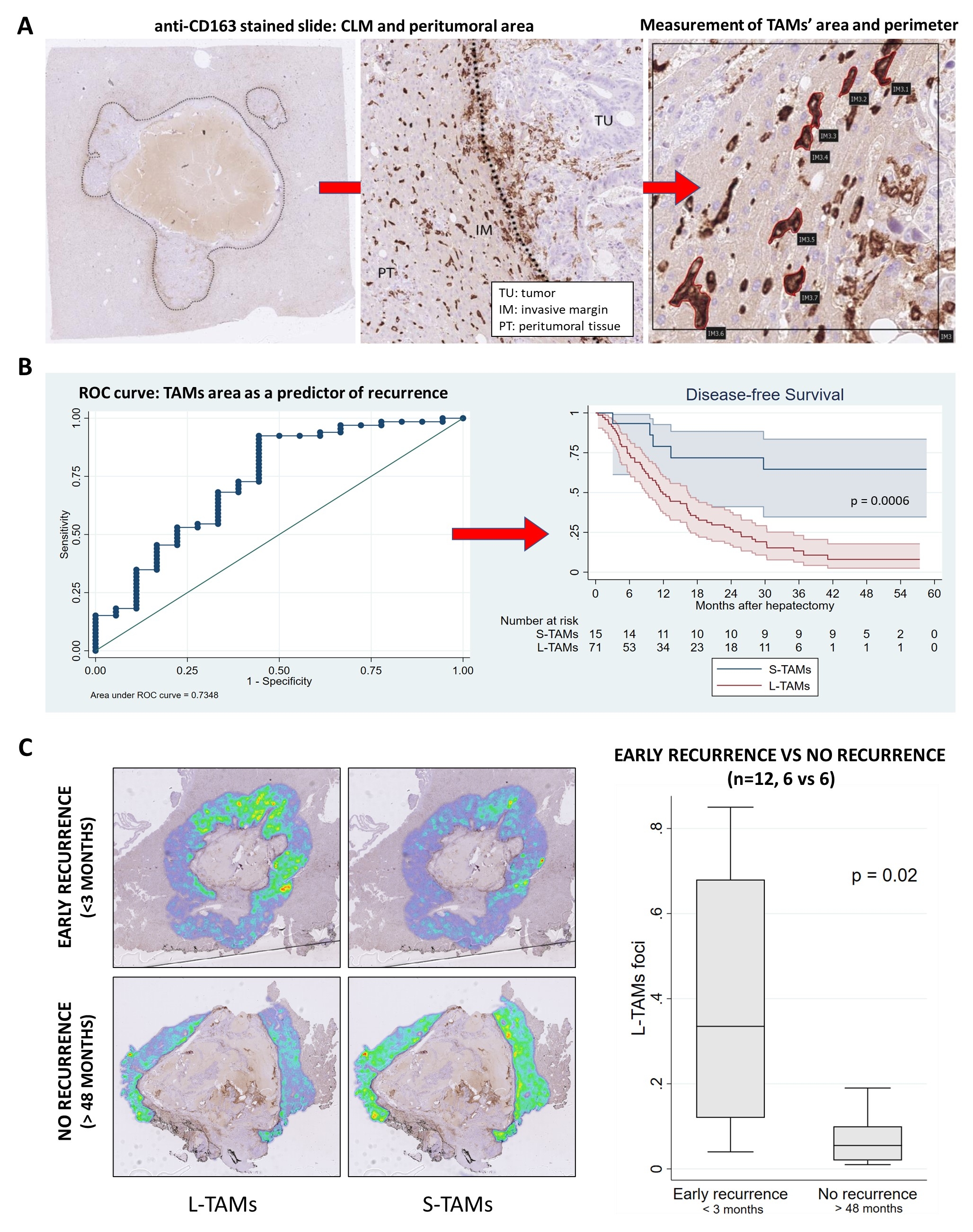
A. Methods, digital pathology
A pathologist blinded to the study, selected three non-contiguous, non-overlapping microscopic (216x216 um2) areas in the invasive margin. In each area, perimeter and area of 7 randomly selected macrophages were calculated by manually tracing cell outlines.
B. Results, TAMs area as a predictor of survival
At univariate analysis, TAM area resulted as an accurate predictor of recurrence (AUC 0.7348) and showed a statistically significant association with DFS (p=0.0006).
C. Result, L-TAMs foci and association with survival
In a subgroup of patients (n=12) that showed early recurrence (< 3 months, n=6) o prolonged disease free survival (>48 months, n=6), foci of L-TAMs were more common in patients showing a less favorable prognosis.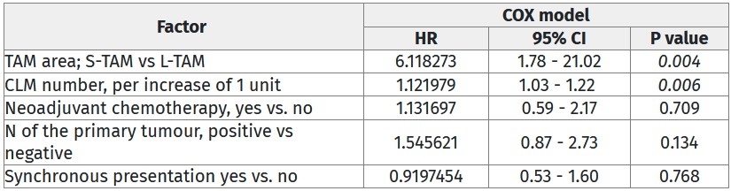
Multivariate analysis result of Cox Regression analysis of prognostic factors for DFS
Back to 2022 Abstracts