
|
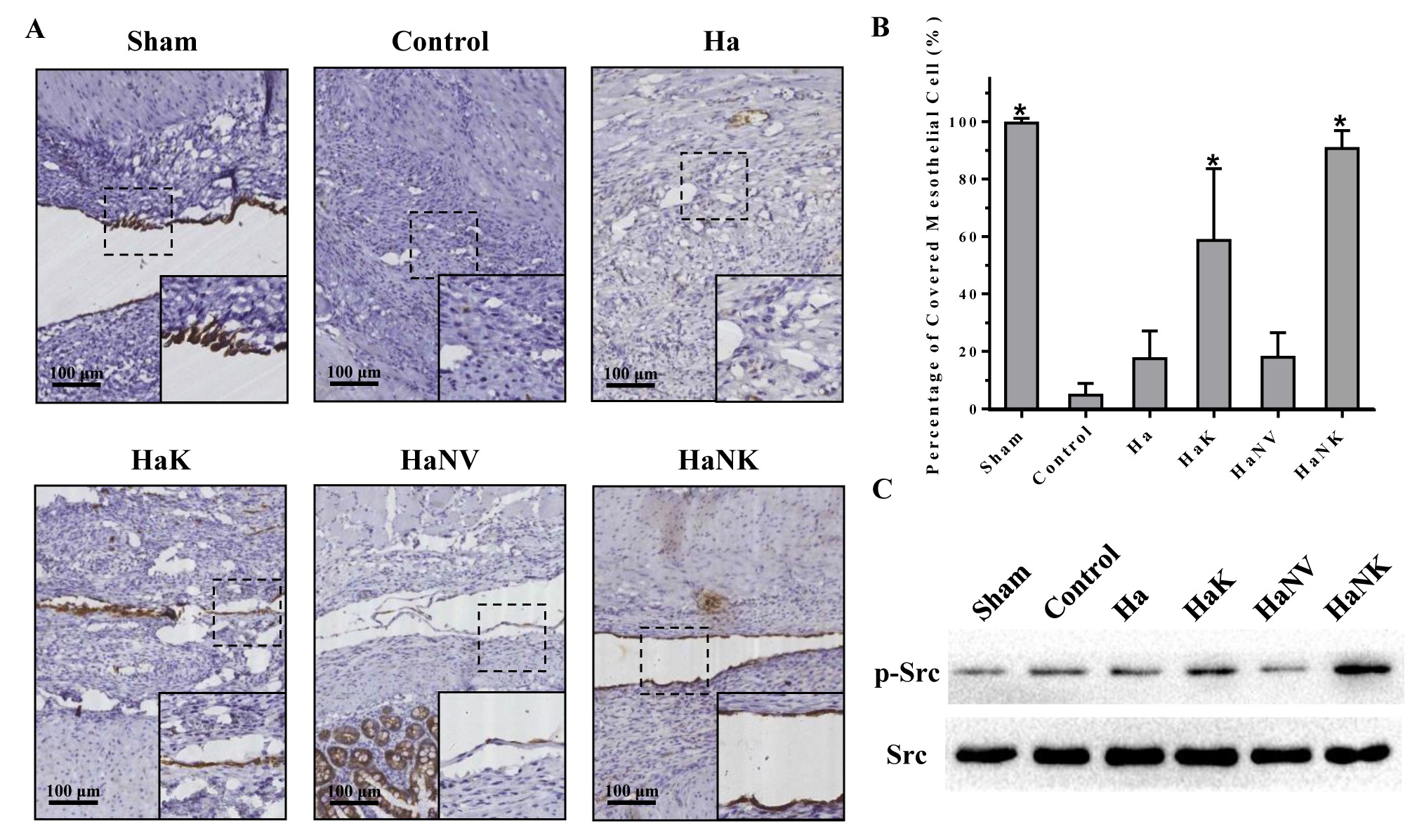
ENGINEERED SELF-ASSEMBLING DOPAMINE-KERATINOCYTE GROWTH FACTOR COLLOIDAL NANOPARTICLES AS ANTI-ADHESION AGENT FOR DETERRING THE POSTOPERATIVE INTRA-ABDOMINAL ADHESIONS Xuqi Li*, Guangbing Wei Department of General Surgery, The First Affiliated Hospital of Xi'an Jiaotong University, Xi'an, Shaanxi, China Background and Aim Postoperative intra-abdominal adhesion (PIAA) is a universal complication that frequently occurs in 90-95% of patients who undergo abdominal surgery. Despite considerable research toward PIAA, the results are still not satisfying with respect to clinical outcome. Thus, developing a simple, facile and economic method to prevent PIAA would be most desirable from both fundamental and practical points of view. Here we report a first attempt to fabricate multifunctional polydopamine-KGF hybrid nanoparticles (PDA-KGF nanoparticles) via a self-assembly strategy, which can be further utilized for preventing the PIAA in the surgical operation. The co-assembling of the dopamine and KGF was tenderly achieved by in situ polymerization technology. Methods Polydopamine nanoparticles were prepared by the oxidative polymerization of dopamine in alkaline water-ethanol solution. KGF as an amine-rich polypeptide can react with dopamine through Michael addition or Schiff base reaction between amine and catechol groups. The cecum wall and its opposite parietal peritoneum were abraded after laparotomy to induce intra-abdominal adhesion formation in rat model. Animals were randomly allocated to receive topical application of different agents. On postoperative day 7, the adhesion score was assessed with a visual scoring system. Tissues were cut off for other assays. Results PDA-KGF nanoparticles were synthesized by the oxidation and self-polymerization of dopamine with KGF in a mixture containing water, ethanol, and tris HCl buffer at room temperature. Typical scanning electron microscopy and transmission electron microscopy imaging revealed that the resultant PDA and PDA-KGF nanoparticles were spherical in shape, with an average diameter of approximately 160 nm. PDA-KGF nanoparticles in combination with Hyaluronate Gel strongly prevented the occurrence of peritoneal adhesions (Fig 1). Specifically, PDA-KGF nanoparticles in combination with Hyaluronate Gel significantly promoted the regeneration and repair of mesothelial cells on the surface of the injured peritoneum (Fig 2). The results indicated that PDA-KGF nanoparticles in combination with Hyaluronate Gel reduced collagen deposition and inhibited fibrosis most significantly during the adhesion formation processes. Conclusion The results from this study showed that PDA-KGF nanoparticles more effectively prevented the formation of PIAA as a component of Hyaluronate and KGF combination therapy. Specifically, the PDA-KGF nanoparticles exhibited significant advantages in promoting mesothelial regeneration, inhibiting inflammatory reaction, and reducing collagen deposition and fibrosis. This improved prophylactic effect is associated with the prolonged activity of PDA-KGF nanoparticles, which persistently promoted the repair of the mesothelial cell layer in the injured peritoneum. 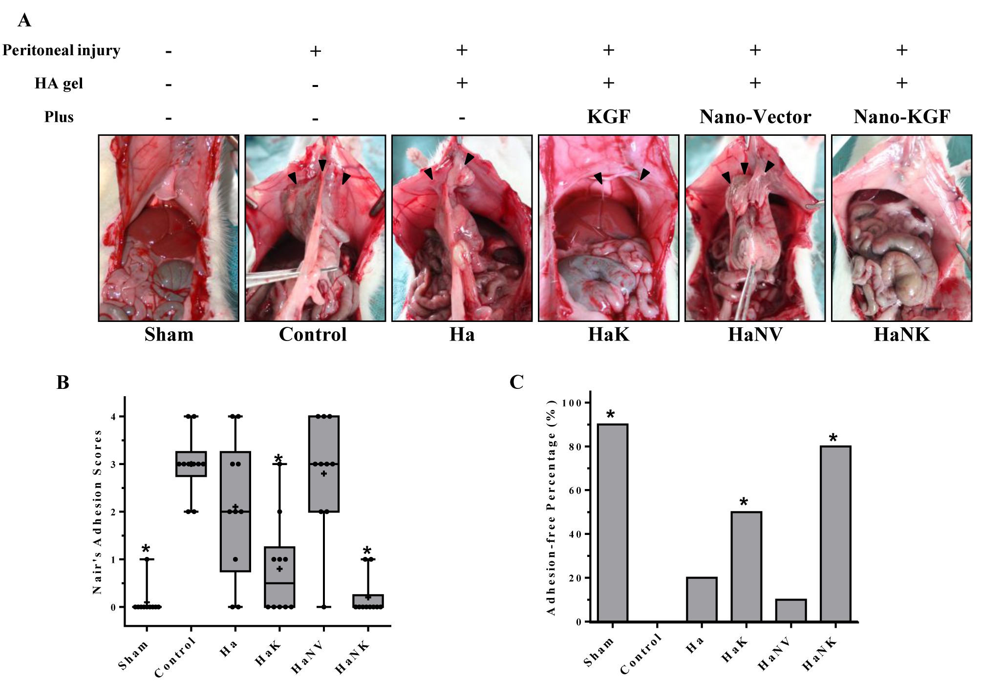
Figure 1. The combined administration of PDA-KGF nanoparticles and Ha prevented postoperative adhesion formation in rats. Back to 2018 Posters |
|||||||||||||||
© 2026 Society for Surgery of the Alimentary Tract. All Rights Reserved. Read the Privacy Policy.