
|
|
Back to 2018 Program and Abstracts
INTRAOPERATIVE MOLECULAR IMAGING WITH A TUMOR HYPOXIA TARGETED NEAR INFRARED CONTRAST AGENT IDENTIFIES SUBCENTIMETER TUMOR DEPOSITS IN A MURINE COLORECTAL CARCINOMATOSIS MODEL
Andrew D. Newton*1, Jarrod D. Predina1, Christopher J. Corbett1, Michael H. Shin1, Lydia G. Frenzel Sulyok1, Michael Baldassari1, Philip S. Low2, Sunil Singhal1
1Surgery, University of Pennsylvania, Perelman School of Medicine, Philadelphia, PA; 2Chemistry, Institute of Drug Discovery, Purdue University, West Lafayette, IN
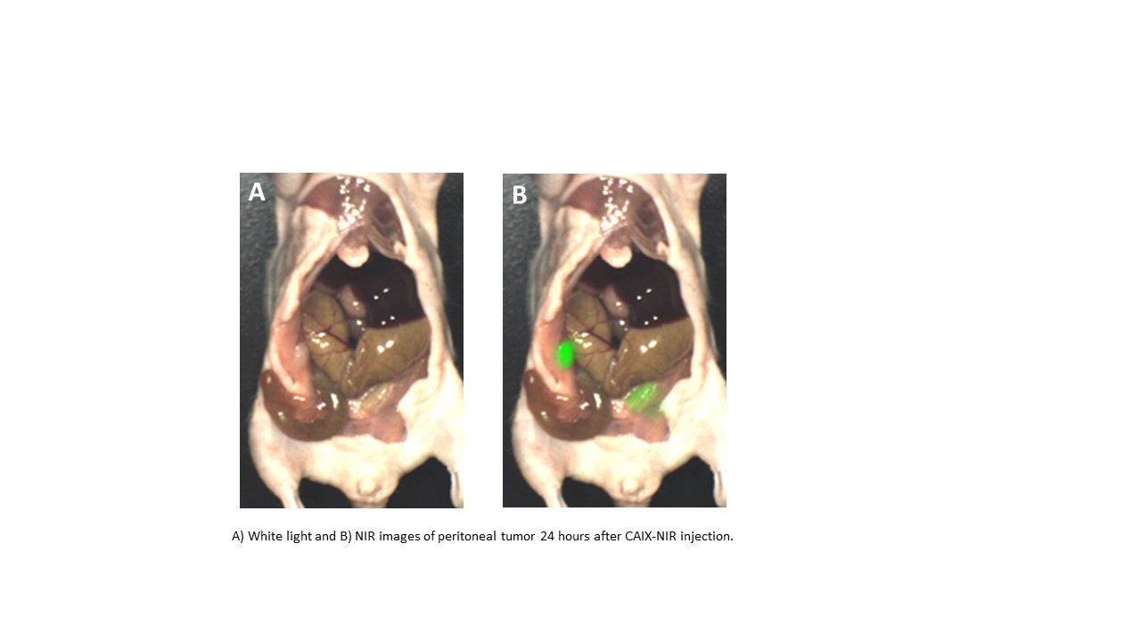
Background: Complete cytoreduction is critical in the surgical treatment of colorectal cancer peritoneal carcinomatosis. The near infrared (NIR) contrast agent indocyanine green (ICG) has previously been used for intraoperative imaging during cytoreduction, but ICG lacks tumor specificity. We hypothesized that intraoperative imaging with a NIR contrast agent targeted to the tissue hypoxia marker carbonic anhydrase IX (CAIX-NIR) would provide improved sensitivity for subcentimeter tumors in a murine model of colorectal cancer carcinomatosis.
Methods: In vitro, CAIX-NIR binding to human colon cancer models (HT29 and HCT116) was confirmed using flow cytometry and fluorescence microscopy. Pharmacokinetics and biodistribution of intravenous CAIX-NIR were determined in mice bearing HT29 and HCT116 xenografts. To compare utility of CAIX-NIR versus ICG, molecular imaging was assessed in an orthotopic model of colorectal carcinomatosis (N=13). Three weeks following intraperitoneal inoculation of HT29 and HCT116 colorectal cancer models, mice received CAIX-NIR or ICG prior to laparotomy. Mice received CAIX-NIR at 0.33 mg/kg 24 hours prior to imaging (N=4), perfusion based ICG at 0.25 mg/kg 30 minutes prior to imaging (N=4), second window ICG at 5 mg/kg 24 hours prior to imaging (N=4), or no dye (N=1). Tumor fluorescence was evaluated using a NIR imaging device currently in human clinical trials (SurgVision® Explorer Air). Tumor specific binding of CAIX was confirmed with fluorescence microscopy.
Results: In vitro, strong fluorescent signal was detected on flow cytometry and fluorescence microscopy after co-incubation of HT29 and HCT116 cell lines with CAIX-NIR. In vivo, peak signal-to-background ratios (SBRs) were seen 16-24 hours after injection of 0.33 mg/kg CAIX-NIR. At 24 hours after injection, the SBR was 50; while strong fluorescent signal was persistent in the stomach and kidneys, weaker signal in the liver and intestine was eliminated. In an orthotopic carcinomatosis model, more fluorescent and total nodules were identified with CAIX-NIR imaging compared to imaging with perfusion based ICG, second window ICG, or no dye (39/43, 0/29, 5/31, and 0/6, respectively, P<0.001). Imaging with CA-IX NIR provided 1 mm sensitivity, while imaging with ICG provided 3 mm sensitivity. Compared to CAIX-NIR imaging, ICG imaging was limited by significant liver and bowel fluorescence. All mice had fluorescence in the stomach and kidneys 24 hours after injection with CAIX-NIR, but it did not interfere with fluorescence from intraperitoneal tumors.
Conclusions: CAIX-NIR is superior to ICG for intraoperative imaging of colorectal cancer peritoneal carcinomatosis in mice. CAIX-NIR is a promising NIR intraoperative imaging agent; it has improved tumor specificity and sensitivity compared to ICG and potential applications in a wide variety of hypoxic tumors.
Back to 2018 Program and Abstracts
|
