|
Back to 2017 Program and Abstracts
PHYSIOLOGY OF ALPPS: LIVER GROWTH AFTER PORTAL VEIN LIGATION CORRELATES WITH THE DEGREE OF NEO-COLLATERALIZATION BETWEEN THE GROWING LOBE AND THE DEPORTALIZED LIVER
Rebecca A. Deal*, Charles Fredericks, Lauren Williams, Edie Y. Chan, Daniel J. Deziel, Martin Hertl, Erik Schadde
General Surgery, Rush University, Chicago, IL
Background: Associating Liver Partition and Portal Vein Ligation for Staged Hepatectomy (ALPPS) induces more rapid liver growth than portal vein ligation (PVL). Our hypothesis is that the transection of liver parenchyma in ALPPS prevents the formation of new portal vein collaterals between lobes. The aim of this study was to determine if prevention of new portal vein collaterals through parenchymal transection impacted growth rates of ALPPS and PVL in pigs.
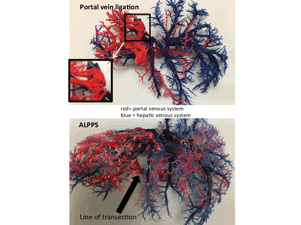
Methods: Models of ALPPS and PVL, using the right posterior lobe as future remnant liver sector, were established in 12 pigs. Animals were randomized to undergo ALPPS (n=4), PVL (n=4) or “partial ALPPS” by varying degrees of transection of the parenchyma (n=4). Hepatic volume was measured by fluid displacement method after 7 days. Portal blood flow and vascular pressure were measured using an ultrasonic flow meter and manometry, respectively. Portal vein collaterals were quantified from epoxy casts.
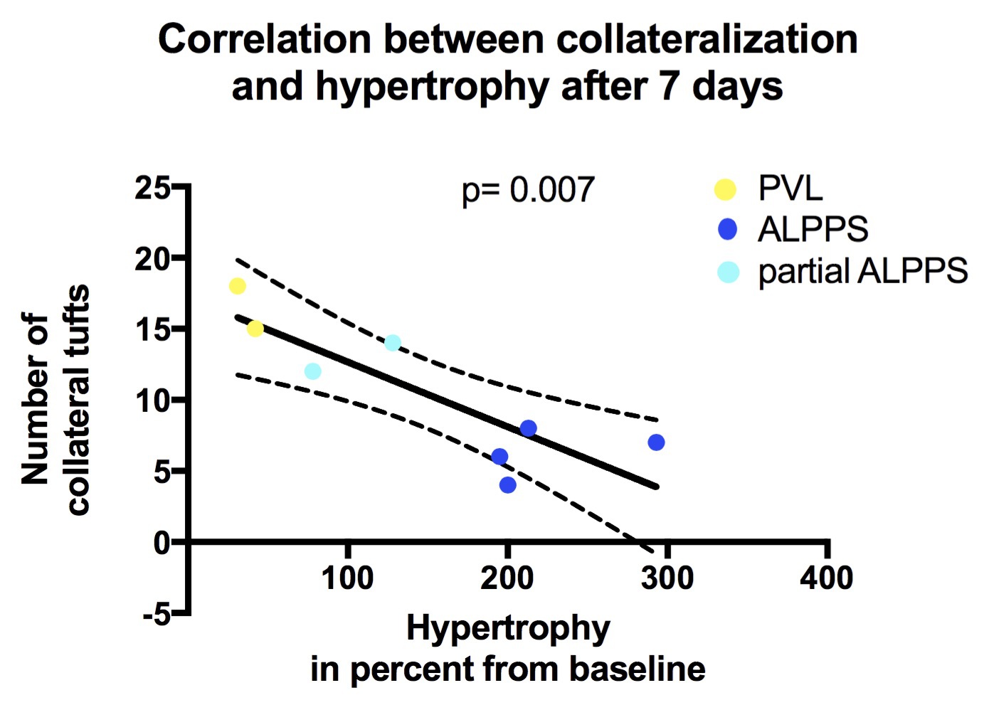
Results: PVL, partial ALPPS, and ALPPS led to differential volume increases of the posterior lobe by 40% (IQR33-45), 128% (IQR78-178), and 207% (IQR196-273) respectively (p<0.005). In PVL and partial ALPPS, substantial new portal vein collaterals were found. The development of collaterals correlated inversely with the growth rate. Portal blood flow was similarly increased for PVL, partial ALPPS and ALPPS.
Conclusions: These data suggest that liver hypertrophy following PVL is inversely proportional to neo-collateralization of the portal vein during the first week. Accelerated hypertrophy after ALPPS is likely due to marked reduction of portal collaterals through transection.


Back to 2017 Program and Abstracts
|


