|
 |
Back to 2017 Program and Abstracts
YTTRIUM-90-LABELED ANTI-GLYPICAN-3 ANTIBODY REDUCES TUMOR GROWTH IN AN ORTHOTOPIC XENOGRAFT MODEL OF HEPATOCELLULAR CARCINOMA
Andrew D. Ludwig*1, Yongwoo D. Seo1, Donald K. Hamlin2, Holly M. Nguyen3, Matthew M. Yeh4, Raymond S. Yeung1, D S. Wilbur2, James O. Park1
1Surgery, University of Washington, Seattle, WA; 2Radiation Oncology, University of Washington, Seattle, WA; 3Urology, University of Washington, Seattle, WA; 4Pathology, University of Washington, Seattle, WA
Background: Hepatocellular carcinoma (HCC) is the second most lethal malignancy and is increasing in incidence in the United States. Unfortunately there are few systemic treatment options, particularly for disseminated disease. Glypican-3 (GPC3) is a proteoglycan cell surface receptor overexpressed in most HCCs and provides a unique target for molecular therapies. We have previously demonstrated that PET imaging using a Zirconium-89-conjugated monoclonal anti-GPC3 antibody can bind to minute tumors and allow imaging with high sensitivity and specificity in an orthotopic xenograft mouse model of HCC and that serum alpha-fetoprotein (AFP) levels are highly correlated with tumor size in this model. In the present study, we conjugated Yttrium-90, a high-energy beta-particle-emitting radionuclide, to our α-GPC3 antibody to develop a novel antibody-directed radiotherapeutic approach for HCC.
Methods: Luciferase-expressing HepG2 human hepatoblastoma cells were injected into the left lobe of the liver in athymic nude mice. At 6 weeks after implantation, tumor establishment was verified by imaging and serum AFP levels from tumor-bearing animals were drawn. Our α-GPC3 antibody was labeled with 90Y using the ligand 1,4,7,10-tetraazacyclododecane-1,4,7,10-tetraacetic acid (DOTA) and injected via tail vein into the experimental mice at a dose of 200 µCi/mouse (n=9) or 300 µCi/mouse (n=9). Control mice received DOTA-conjugated antibody without radionuclide (n=7). Serum AFP levels were drawn at 14 and 30 days after treatment. The animals were then sacrificed and the livers were removed en bloc and weighed. Immunohistochemistry was performed on the tumors to confirm antibody delivery and evaluate the effect of radiation treatment.
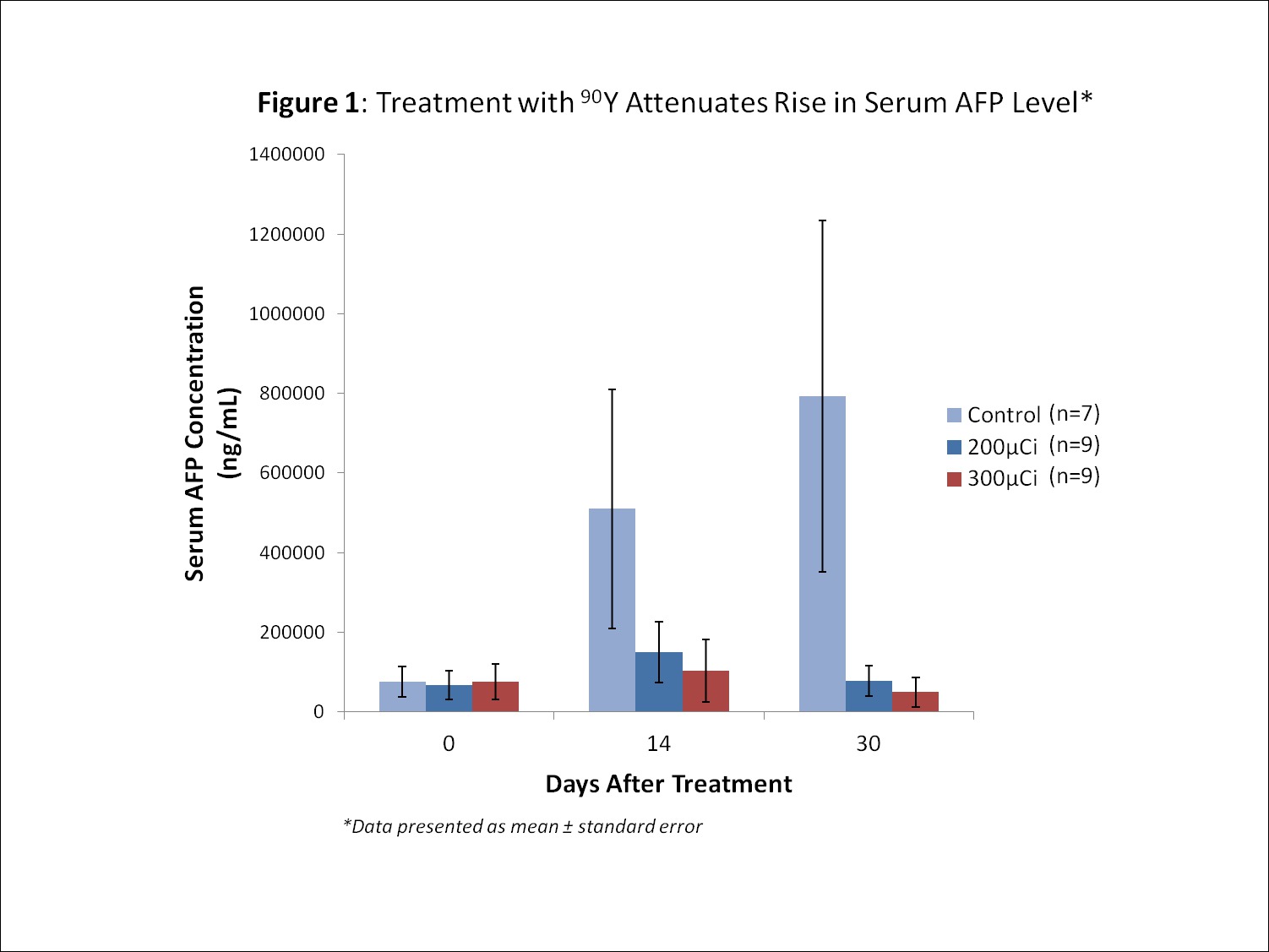
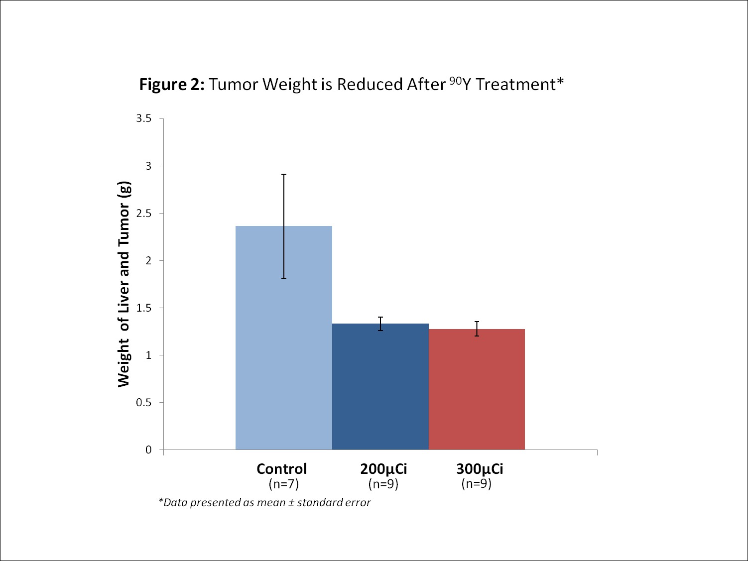
Results: Before treatment there was no statistical difference between serum AFP levels or tumor size among any of the three groups. Mean serum AFP levels in control animals increased by 663% over 30 days, while animals treated with 200 µCi 90Y experienced a 28% increase and animals treated with 300 µCi had a 42% reduction in mean serum AFP (p=0.03 and 0.02, respectively), [Fig. 1]. Mean liver weight in control animals reached 2.36g compared to 1.33g in animals that received 200 µCi 90Y and 1.28g in animals that received 300 µCi (p=0.05 and 0.04, respectively), [Fig. 2]. These results were achieved without significant toxicity as measured by body condition scoring and body weight.
Conclusions: The results of this pilot experiment demonstrate that GPC3, a cell-surface proteoglycan overexpressed in most HCC, can be used as a target for radioimmunotherapy in an orthotopic mouse model of HCC. These data suggest that this novel targeted approach could be used in the clinical setting, particularly for disseminated HCC.


Back to 2017 Program and Abstracts
|


