|


|
Back to 2015 Annual Meeting Program Trisulfated Disaccharide Decreases Intracellular Calcium Concentration in Hepatocytes Under Calcium Overload Situation ÊNio R. Vasques*1, José Eduardo M. Cunha1, ANA Maria M. Coelho1, Sandra N. Sampietre1, Eleazar Chaib1, Helena B. Nader2, Ivarne S. Tersariol2, Luiz C. D Albuquerque1, Tiago Rodrigues3 1GASTROENTEROLOGY, FMUSP, São Paulo, Brazil; 2UNIFESP, São Paulo, Brazil; 3UFABC, Santo Andre, Brazil
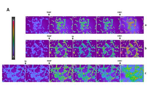
Background: Ischemia and reperfusion (I/R) with intracellular calcium overload are pivotal phenomena in tissue damage in liver surgery and transplantation. Previous studies in our laboratory showed the Trisulfated Disaccharide (TD) with 585 Da molecular weight has no effects on coagulation mechanisms and protects hepatocytes from I/R-induced injury in rats. The sodium/calcium exchanger (NCX) that regulates calcium extrusion is affected by TD in endothelial aortic cells in rabbits by decreasing intracellular calcium through the inhibition of the exchange inhibitory peptide (XIP), however, the mechanism involved in liver protection is unclear. Objective: To evaluate the TD effect in vitro in hepatocytes under calcium overload. Material and Methods: BRL3A culture cells from Rattus norvegicus liver were grown in supplemented Dulbecco's modified Eagle's medium for 24h. Then, cells were loaded with the fluorescent Ca2+ indicator fura-2/AM (4 μM) for 30 minutes at 37°C in a 5% CO2 atmosphere. After washing twice, 1.0 mL of medium containing 1.5 mM CaCl2, 130 mM NaCl, 5.6 mM KCl, 0.8 mM MgSO4, 1 mM Na2HPO4, 25 mM glucose, 2 mM HEPES, and 2.5 mM NaHCO3 (pH 7.3) were add to cells and the images were acquired on a widefield microscope coupled to a digital camera and external filter wheel. Changes in cytosolic calcium levels were measured kinetically at 37°C for 15 minutes using excitation at 340 and 380 nm simultaneously, and emission captured at 505 nm. The ratio images and curves were obtained using the calcium imaging mode in Leica Application Suite software. It was used 200 µM thapsigargin to obtain a sustained raise in the cytosolic calcium level. To evaluate TD effect on calcium homeostasis, 30 µM TD was added to the medium prior and after thapsigargin (n=3). Results: TD added to the medium after thapsigargin decreased transiently the intracellular calcium levels and its effect exhibited duration of about 250 seconds. Although TD addition prior thapsigargin resulted in a slight decrease in the intracellular basal calcium levels, it decreased significantly the extent of the thapsigargin-induced cytosolic calcium increase (fig. 1A, 1B). Conclusion: TD was able to decrease the intracellular calcium raise promoted by thapsigargin and to prevent it when added prior to the inducer in liver culture cells. Back to 2015 Annual Meeting Program |
|||||||||
© 2026 Society for Surgery of the Alimentary Tract. All Rights Reserved. Read the Privacy Policy.