|
Back to 2015 Annual Meeting Program
Laparoscopic Surgery for Median Arcuate Ligament Syndrome (MALS): Report of First Hybrid Intraoperative Angiography Case and a Review of the Literature
Gowri Kabbur*1, 2, James Brockett1, David Williams1, Robert Netzley1, Charu Paranjape1
1Surgery, Akron General Medical Center, Akron, OH; 2Northeast Ohio Medical University, Rootstown, OH
Introduction: Median arcuate ligament syndrome (MALS) is a relatively rare condition caused by compression of the celiac trunk, most commonly from low lying diaphragmatic crural fibers. Patients typically present with postprandial abdominal pain and weight loss secondary to "food fear". Advanced laparoscopic approaches have gained popularity over the past decade due to decreased complication rates and postoperative hospital stay. Our particular interest lies in the use of intraoperative angiography imaging before and after the laparoscopic release of the median arcuate ligament to acutely assess decompression of the celiac trunk.
Methods: We performed a retrospective literature review on PubMed for all case reports published on laparoscopic MALS in the adult population. Criteria for inclusion in the review included adults aged 19+, a laparoscopic operative approach, and publication in an English language journal. Fifteen case reports met criteria (ours being the sixteenth) and a detailed comparative data analysis was performed for each article, with focus placed on preoperative diagnosis, intraoperative procedures, postoperative diagnosis, resolution of symptoms, and recurrence of celiac stenosis.
Findings: The results of our literature review are outlined in Table 1. A total of 79 laparoscopic MALS cases were reported in 16 case reports published between 2000-2014 (including our own case). The patient population was predominantly female (n=62, 78%), with a mean reported BMI of 19.2. The most favored mode of preoperative imaging used was CT angiography (n=51, 65%), closely followed by Duplex Doppler ultrasonography (n=48, 61%). There were 22 reported intraoperative complications, with 8 conversions to open laparotomy. Only 6 laparoscopic cases used intraoperative confirmatory procedures, the majority being Doppler ultrasonography (80%) and ours being the FIRST reported intraoperative hybrid angiography. 66 patients (84%) reported complete resolution of symptoms (including our own patient), 7 patients reported partial resolution (9%), and 5 patients had no resolution of symptoms (6%).
Conclusion: A laparoscopic surgical approach for median arcuate ligament syndrome can be performed with low morbidity and mortality and has shown to have significant benefits such as shorter operative time, decreased hospital stay, and lower recurrence of symptoms. Our literature review demonstrates a lack of reported use of intraoperative confirmatory imaging during laparoscopic release of the median arcuate ligament. We report the FIRST CASE of median arcuate ligament syndrome treated laparoscopically with intraoperative hybrid angiography. Use of intraoperative confirmatory tests such as Duplex doppler and angiography may result in higher rates of complete symptom resolution.
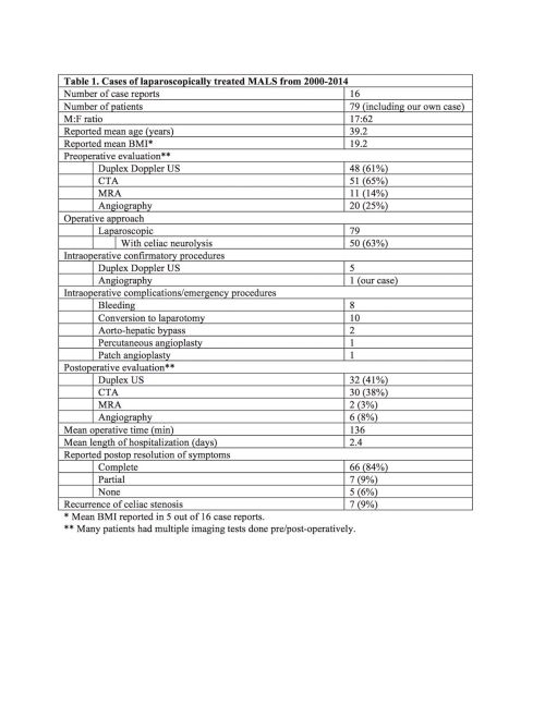
Table 1
Back to 2015 Annual Meeting Program
|

