|
Back to 2015 Annual Meeting Program
Laparoscopic Treatment of a Giant Colonic Lipoma Presenting As Transverse Colon Intussusception and Lower Intestinal Bleeding: a Case Report
Beatriz De Rienzo*, Horacio Montanez-Ramirez, Fernando Quijano-Orvananos
American British Cowdray Medical Center, Alvaro Obregon, Mexico
Introduction: Intussusception in adults accounts for 5% of bowel intussusceptions and of 1% of bowel obstructions. Most adult intussusceptions have a demonstrable lead point. Colonic lipomas are a rare cause of intussusception. They are usually small and asymptomatic, with only 30% reaching more than 2 cm at which point they develop symptoms which include anemia, abdominal pain, constipation, diarrhea, bleeding and intussusception. Giant lipoma has been defined as >5 cm in diameter. We present a case of a 6 cm lipoma in the transverse colon causing gastrointestinal bleeding and colo-colonic intussusception.
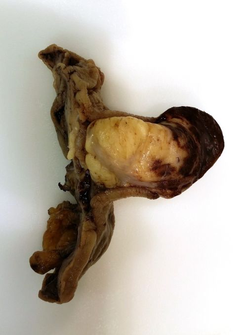
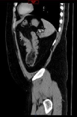
Case: A 31 year-old male patient presented to the emergency room with a one month history of melena and hematochezia, colicky abdominal pain and in the previous 24 hours nausea, vomiting and abdominal distention. Contrast-enhanced CT scan showed a colo-colonic invagination of transverse colon telescoping into the descending colon (Fig. 1). An enema with hidrosoluble contrast was performed with resolution of the intussusception, and an intraluminal oval pediculated mass of 65x41x37 mm with a density of -21 HU was observed at the splenic angle. Colonoscopy reported a submucosal mass with a blood clot causing obstruction of the bowel lumen 55 cm from the anal verge; its proximal and distal margins were marked with blue dye. The patient was taken to surgery and a laparoscopic segmental transverse colectomy was performed. The histopathological report revealed a 6x5x4 cm submucosal lipoma (Fig. 2). The postoperative evolution was uneventful and the patient was discharged on the fifth postoperative day.
Discussion: Colonic lipomas are benign non-epithelial tumors which present most commonly in females within the fifth and sixth decades of life. They are most commonly located in the ascending colon (61%), followed by the descending colon (20%), transverse colon (15%) and rectum (3.4%). Symptoms appear in lipomas >2 cm: anemia, colicky abdominal pain, constipation, bleeding and intussusception. Diagnostic methods include CT scan, contrast-enhanced enema, and colonoscopy. CT scan will reveal a regular, well-delimited spherical or ovoid mass of adipose density (-40 to -120 HU). If associated with intussusception it will show a target lesion, a sausage-shaped mass with layers or a mass caused by edema, mural thickening and vascular compromise. Contrast enhanced enema may aid in the reduction of an intussusception and additionally demonstrate a filling defect. Colonoscopy will reveal a submucosal lesion with three characteristic signs: "cushion sign," "tenting effect," and "naked fat sign." Treatment for symptomatic lipomas includes endoscopic excision or surgical resection, with size being the delimiting factor. Segmental colectomy is the gold standard and may be performed laparoscopically.
Back to 2015 Annual Meeting Program
|

