SSAT Home SSAT Annual Meeting

Back to SSAT Site
Annual Meeting Home
Past & Future Meetings
Photo Gallery
 

Back to 2015 Annual Meeting Program


Depletion of Enteric Microbiota Protects from Chemotherapy-induced Damage in the Murine Small Intestine
Jacquelyn Carr*, Stephanie L. King, Christopher M. Dekaney
Surgery, UNC, Chapel Hill, North Carolina

Introduction: Enteritis is a significant side effect of chemotherapy, and is a potential dose-limiting complication. Pharmacotherapy can diminish symptoms, but there are no effective treatments to prevent the intestinal epithelial injury responsible for enteritis. While the intestinal epithelium can repair and regenerate, the mechanisms guiding epithelial damage and repair are unknown. Our preliminary studies suggest that enteric bacteria play a critical role in precipitating mucosal damage after treatment with doxorubicin (Dox), a common chemotherapeutic drug. Following Dox, mice raised in germ free conditions do not show the small intestine crypt loss, increased crypt depth, or alterations in proliferation demonstrated in conventionally raised mice. In the germ free model, the mice have never been exposed to bacteria, which naturally alters their enteric flora. Since the enteric bacterial environment is crucial for homeostasis, this complete lack of exposure to bacteria likely alters intestinal signaling not only at baseline, but also in response to damage. In the current study, we thus employ a model to eliminate enteric bacteria in adult mice in order to more fully elucidate the role of bacterial signaling in moderating small intestinal damage to Dox.

Methods: At 10 weeks of age C57/BL6 mice were treated with or without a panel of antibiotics (vancomycin 500 mg/L, ampicillin 1g/L, neomycin 1g/L, metronidazole 1g/L) in drinking water for 4 weeks. Depletion of bacterial flora in antibiotic-treated (ABRX) mice was confirmed by culture of feces at three and four weeks. After 4 weeks, mice were treated with Dox (20mg/kg) for 0 to 5 days. The jejunum was fixed and stained with H&E or anti-phosphohistone H3 for histologic analysis.

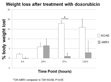
Results: ABRX mice lost less weight at 72 and 120 hours following Dox treatment than mice that did not receive antibiotics (NOAB) (graph). ABRX mice demonstrated a baseline increase in circumference following antibiotic treatment, compared to NOAB mice at baseline. ABRX mice showed significantly greater crypt survival, shallower crypts, and fewer proliferating cells on phosphohistone H3 staining at 120 hours after Dox than NOAB mice (table).

Conclusions: Adult mice whose enteric flora has been depleted undergo an adaptive response in which they expand the circumference of their jejunum by approximately 25%. In response to insult with the chemotherapeutic Dox, these mice demonstrate significantly less intestinal damage than mice that were not treated with antibiotics. They also do not show the deepening of crypts or increase in crypt proliferation typical of the repair process in non-antibiotic treated mice. Future directions include investigation of the specific bacterial signaling pathways responsible for this difference in order to determine a molecular target for prevention of chemotherapy-induced enteritis.


Back to 2015 Annual Meeting Program


© 2024 Society for Surgery of the Alimentary Tract. All Rights Reserved. Read the Privacy Policy.