|
Back to 2014 Annual Meeting Posters
MRI Liver in Pancreatic Cancer: a Game Changer
Cindy Chew*1, Hedvig Karteszi1, Nigel B. Jamieson2, Ross Carter2, Euan J. Dickson2, Colin Mckay2
1Radiology, Glasgow Royal Infirmary, Glasgow, United Kingdom; 2Surgery, Glasgow Royal Infirmary, Glasgow, United Kingdom
Background: Pancreatic cancer is one of the leading causes of cancer mortality. Surgery is the only chance of cure, but is inappropriate for patients with metastasis.
Aim: To evaluate the frequency of liver metastasis on MRI in patients with resectable pancreatic cancer and normal liver on contrast enhanced MDCT.
Methods: Between April 2012-13, all patients with resectable pancreatic cancer based on CT staging underwent MRI liver - utilising hepatocyte specific contrast agent and diffusion weight imaging.
Results: Forty five consecutive patients were examined. Thirty two were male and 13 were female. Median age was 64 years (range 31-76 years). MRI was performed at a median of 2 weeks from CT. Thirteen (29%) patients with normal liver on CT had findings consistent with liver metastases while 4 (9%) had indeterminate liver lesions on MRI. Three of the 4 patients with indeterminate liver lesions demonstrated progression on follow up imaging consistent with metastases. One of the 28 patients with a normal MRI liver underwent palliative bypass on discovering a 5mm subcapsular lesion at laparotomy. At a median follow up of 12 months, 3 of the remaining 27 patients with normal MRI had evidence of liver metastasis (7, 10 and13 months post MRI) while 4 had extrahepatic disease. Survival was significantly reduced in patients with liver metastasis on MRI (p=0.01).
Conclusion: MRI identified liver metastases in 36% of patients with resectable pancreatic cancer on MDCT and should be included in the routine staging of these patients.
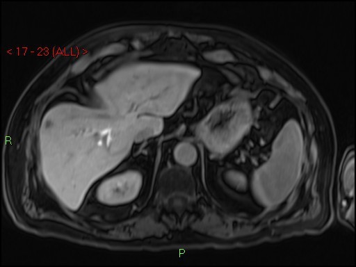
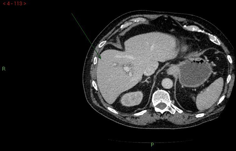
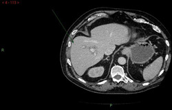 Normal liver staging CT. 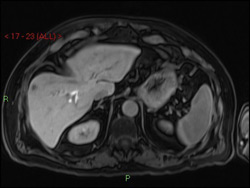 One of multiple lesions seen on MRI consistent with multiple liver metastases (subcapsular location, segment VIII). Follow up CT 4 months later demonstrated increase in the number and size of liver metastases.
Back to 2014 Annual Meeting Posters
|