
|
|
Back to Annual Meeting Posters
The Inflammatory Microenvironment in Duodeno-Esophageal Reflux Induced Esophageal Carcinogenesis in a Sequential Rat Model
Tomoharu Miyashita*1,2, Masayoshi Munemoto1, Furhawn a. Shah2, John W. Harmon2, Takashi Fujimura1, Daisuke Matsui1, Masanobu Oshima3, Tetsuo Ohta1
1Department of Gastroenterologic Surgery, Kanazawa University Hospital, Kanazawa, Japan; 2Department of Surgery, Johns Hopkins Bayview Medical Center, Baltimore, MD; 3Division of Genetics, Cancer Research Institute, Kanazawa University, Kanazawa, Japan
Background: Macrophages play an important role in tumorigenesis. Macrophages are polarized to either the classical M1 type or alternative M2 type. Tumor-associated macrophages (TAMs) are polarized to M2 or M2-like types and have been shown to promote the progression and metastasis of cancer. We hypothesized that TAMs in an inflammatory microenvironment induced by duodenal content reflux without carcinogens may promote the development of esophageal carcinomas.
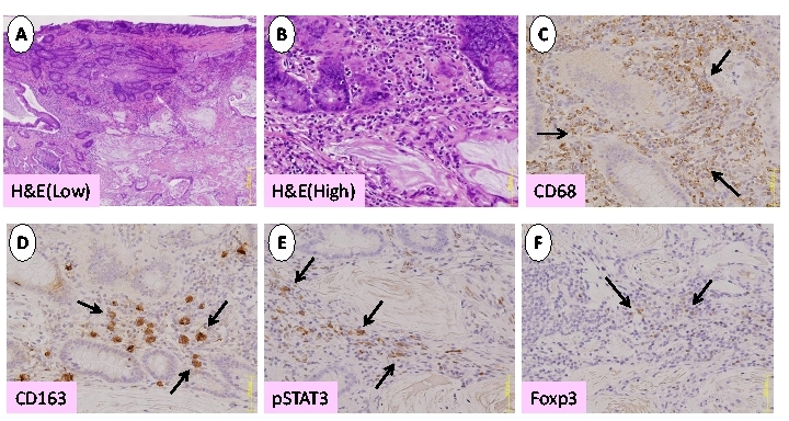
Methods: A total gastrectomy followed by esophago-jejunostomy was performed on rats in order to induce chronic duodenal content reflux esophagitis. The animals were sacrificed sequentially, at the 20th, 30th and 40th week after surgery and their esophagi were examined. Primary antibodies against CD68(pan-macrophage; BMA Bio), CD163(M2; AbD Serotec), pStat3(Cell Signaling), Foxp3(Treg; eBioscience) were used to evaluate the expression and localization of the inflammatory response.
Results: At 20-30 weeks post-surgery, squamous proliferative hyperplasia (PHP) and Barrett's metaplasia (BM) were observed. Adenocarcinoma (ADC) associated BM (Fig1A,B) and squamous cell carcinoma (SCC) were observed 40 weeks post-surgery. Numerous CD68 positive macrophages were identified surrounding PHP and BM at 20 weeks and surrounding ADC and SCC after 40 weeks (Fig1C). In contrast, few CD163 positive macrophages infiltrated into the PHP, BM, ADC and SCC after 40 weeks (Fig1D). The PHP, BM, ADC and SCC lesions exhibited some pStat3-positive cells (Fig1E). A few Foxp3-positive cells were detected near carcinoma lesions after 40 weeks (Fig1F).
Conclusions:Our data showed that macrophages infiltrate the esophagus at the early inflammatory stage of carcinogenesis. In the inflammatory microenvironment, characteristic of the activated pStat3 pathway, M2 phenotype macrophages infiltrate and contribute to tumor development. Furthermore, Treg was induced by tumor relating to process of carcinogenesis
Back to Annual Meeting Posters
|

