|
Back to Annual Meeting Program
Molecular Predictors of Recurrent Hepatocellular Cancer: Role of Cancer Stem Cells
Prejesh Philips*, Xuanyi LI, Yan LI, Suping LI, Erik M. Dunki-Jacobs, Robert C. Martin
Surgical Oncology, University of Louisville, Louisville, KY
Background: Recurrence rates after either resection of hepatocellular carcinoma (HCC) or liver transplantation occur in 25 to 75% of patients. HCC recurrence has been thought to be driven by cancer stem cells (CSCs). Understanding the role CSCs play in HCC recurrence will provide the important information to improve prognosis and better define adjuvant therapy.
Aim: To demonstrate that HCC cells can dedifferentiate into CSCs, which contribute to HCC is an important resource.
Method: The capacity of CSC sphere formation was tested in a human HCC cell line, Hep3B (derived from an 8-year-old black patient). The CSC sphere condition medium was the DMEM/ F12 medium (1:1) supplied with 20 ng/ml of EGF and 20 ng/ml of bFGF. Flow cytometery were performed using CSCs surface antigens (including, CD133, CD90, CD44, and EpCAM). To test the ability of CSCs for tumor formation, an orthotopic model was developed using nude BALB-B/C. Tumor inoculation were performed using Hep3B cells and Hep3B CSCs at 2x10⁶ per injection. The mice were assessed for tumor formation at 4 weeks. Flow cytometery were performed in the cells isolated from tumor tissue to test CSCs surface antigens mentioned above.
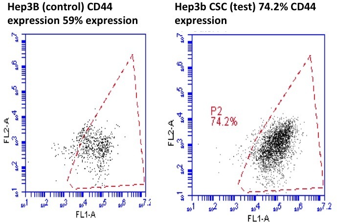
Results: Sphere forming Hep3B cells were successfully induced and confirmed to contain CSCs morphology. Compared to the regular cultured Hep3B cells, Hep3B sphere cells demonstrated significantly higher surface antigens. CD44 was expressed by 74.25% of CSC vs. 59.79% regular Hep3B cells (difference of 24.46%, p value=0.029). With regards to EPCAM expression the CSC cells expressed 1.6% versus 0.51% expressed by regular Hep 3b cells (p value=0.02). This enhanced expression dropped down to near baseline when the Hep3B sphere re-cultured in standard nutrient rich medium. CD44 and EPCAM expression was noted at 60.1% and 0.6%, which was not significantly different compared to regular Hep3b cells (p=0.87 and 0.8) but was significantly lower compared to CSC cells (p=0.03 and 0.0.05). In the orthotpoic injection liver, Hep3B sphere demonstrated a significantly higher tumor proliferation rate compared to non-sphere Hep3B cells. The tumor weights are as follows: 389mg±65 (Hep3B sphere) vs 94mg±32 (Hep3B cells).
Conclusions: Hep3B cells show the capacity of CSC induction in nutritionally stressed phase. Hep3B derived CSC can not only differentiate into HCC cells when supplied with nutrient rich medium, but also form tumor when inoculate into mouse liver. The study is on the way to investigate the signaling such as Wnt pathway to evaluate the clinical relevant biomarkers of CSCs in HCC patients.

Hep3B expression of CD 44 (sphere forming) left (FITC +ve 74.25%) versus control Hep3B cells right (59.79%)
Back to Annual Meeting Program
|


