|
|
Back to Annual Meeting Program In Rats After Esophagojejunostomy, Reflux Esophagitis Is Accompanied by the Expression of SOX-9 in Basal Cells of the Squamous Epithelium and in Barrett's Metaplasia Thai H. Pham*, David H. Wang, Robert M. Genta, Shelby D. Melton, Chunhua Yu, Stuart J. Spechler, Rhonda F. Souza, William Neumann Surgery, North Texas VAMC; UT Southwestern Medical Center, Dallas, TX
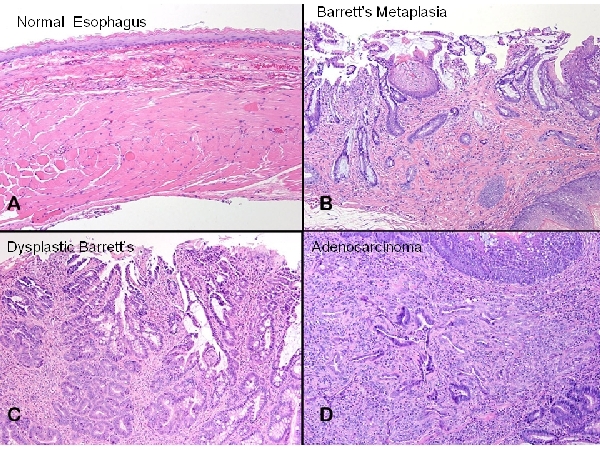
Introduction: Metaplasia involves the change from one adult cell type into another that is phenotypically different, but that is often of similar embryonic origin. The embryonic esophagus initially is lined by columnar cells that are replaced by squamous cells as maturation proceeds. Barrett’s metaplasia involves the change from esophageal squamous cells back into columnar cells in the setting of gastroesophageal reflux disease. SOX-9, a transcription factor that regulates the development of columnar cell morphological features, is expressed in Barrett’s metaplasia and in the mouse embryonic, columnar-lined esophagus, but not in the normal adult squamous-lined esophagus. Furthermore, forced expression of SOX-9 in cultured esophageal squamous cells induces a columnar phenotype. We sought to determine whether SOX-9 expression is involved in the development of Barrett’s metaplasia in rats that have reflux esophagitis induced by esophagojejunostomy (EJ). Methods: Groups of 5 Sprague-Dawley rats were sacrificed at 8, 10, 16, and 24 weeks after EJ. The distal esophagus was removed, sectioned, paraffin-embedded and mounted on slides, which were stained with H&E for histological evaluation; immunohistochemistry was performed to determine SOX-9 protein expression. We evaluated the specimens for 1) squamous basal cell and papillary hyperplasia, 2) Barrett’s metaplasia with and without dysplasia, and 3) adenocarcinoma. SOX-9 expression was assessed only in squamous epithelium and in non-dysplastic Barrett’s metaplasia. Sham-operated animals were used as controls. Results: At 8 weeks after EJ, erosive esophagitis with prominent squamous basal cell and papillary hyperplasia was present in all animals. In addition, some of the squamous cells appeared to produce mucin, which was present both within and between cells. At 8 weeks, non-dysplastic Barrett’s metaplasia, dysplastic Barrett’s metaplasia, and adenocarcinoma were found in 4, 3 and 1 of the 5 rats, respectively (Figure 1B-D). Similar histologic findings were seen at the later time points but not in sham-operated animals (Figure 1A). SOX-9 was expressed by basal cells of the squamous epithelium close to the EJ anastomosis (Figure 2A), but not in squamous epithelium further from the anastomosis. Intense expression of SOX-9 was detected in areas of non-dysplastic Barrett’s metaplasia (Figure 2B). Control animals did not show any esophageal SOX-9 expression. Conclusions: In rats after esophagojejunostomy, the development of reflux esophagitis is accompanied by expression of SOX-9 in the basal cell layer of esophageal squamous epithelium near the anastomosis. In addition, SOX-9 is expressed in Barrett’s metaplasia in this rat model. These data suggest that this is a relevant model for studying the role of SOX-9 in the development of Barrett’s esophagus and esophageal adenocarcinoma. Back to Annual Meeting Program
|
||||||||
© 2026 Society for Surgery of the Alimentary Tract. All Rights Reserved. Read the Privacy Policy.


