
|
|
Back to Annual Meeting Program
Thromboelastography Delineates Hypercoagulation in an Immunocompentent Murine Model of Metastatic Colon Cancer
Karen K. Lo*1,2, Theresa Chin1, Marguerite Kelher3, Martin Mccarter1, Ernest E. Moore2, Christopher Silliman1,3, Carlton C. Barnett2
1Surgery, University of Colorado, Denver, Aurora, CO; 2Surgery, Denver Health, Denver, CO; 3Bonfils Blood Center, Denver, CO
INTRODUCTION: The association between malignancy and venous thrombosis (VTE) has been well documented since the 1860s. Moreover, it has been demonstrated that perioperative blood transfusions increase the risk of VTE in colon cancer patients. Despite efforts to prevent VTE, current diagnostic tests (INR, PTT, PT, platelet count) are often unreliable. Recently, our lab has demonstrated that Thromboelastography (TEG) is able to better assess coagulation kinetics and direct patient therapy than conventional testing. We hypothesize TEG will delineate coagulation abnormalities in a murine model of transfusion mediated metastatic colon cancer.
METHODS: C57/BL6 male mice, age 7-9 weeks, underwent splenic inoculation with 2.5 x 10⁴ MC38 murine colon adenocarcinoma cells. Control mice underwent the same surgery with splenic injection of normal saline. One week after inoculation, all mice were randomized to receive blood transfusion via tail vein injection in the amount of 1 mg/kg or the equivalent dose of normal saline. N ≥ 4 in all groups. Three weeks after cancer inoculation, cardiac puncture was performed and blood was collected with citrate in a 1:10 ratio. TEG was performed on TEG® 5000 Thrombelastograph® Hemostasis Analyzer. Necropsies were then performed: tumors were harvested and metastases were determined. TEG was compared between mice with metastatic cancer with and without transfusion and control mice, who received sham cancer surgery, with and without transfusion. Data were analyzed using ANOVA with p≤0.05 used to determine significance.
RESULTS: Mice with cancer that received blood transfusions were found by TEG to have significantly lower R times (4.4 minutes versus 8.5 minutes p=0.018), K times (1.6 minutes versus 3.3 minutes p=0.0004), and significantly higher angles (67° versus 52° p=0.0005), MA (68 mm versus 62 mm p=0.019), and G (10.7 versus 8.05 dynes/cm2 p=0.04) when compared to mice who received a sham operation and blood transfusions. TEG value interpretation shown (Figure 1); R times demonstrate that mice with metastatic colon cancer form clot significantly faster than mice without cancer (Figure 2). Surprisingly, blood product transfusion did not affect hypercoagulabilty.
CONCLUSION: TEG is able to delineate hypercoagulabilty associated with metastatic colon cancer in an immunocompetent murine model. Receipt of packed red blood cell product did not affect hypercoaguability in this model. As thrombotic events are morbid and potentially mortal, additional investigation of this modality in perioperative management of cancer patients appears warranted.
TEG Value Interpretation
| Value | Meaning | Decrease indicates: | Increase indicates: | | R | Clotting Time, i.e. time (minutes) until the first detectable levels of fibrin clot formation. Generally reflects coagulation factor levels | Hypercoagulable | Hypocoagulable Factor deficiency, anticoagulant, hypofibrinoginemia | | K | Clot Kinetics. Measures the speed to reach clot strength of 20mm amplitude. Looks at intrinsic clotting factors, fibrinogen, platelet function | Hypercoagulable | Hypocoagulable | | Angle | Clot strengthening, rapidity of fibrin-buildup and clot formation, angle of tracing from r to K value. | Hypocoagulable Hypofibrinogenemia or thrombocytopenia | Hypercoagulable | | MA, G | Overall Clot strength, represents maximum dynamics of fibrin and platelet bonding | Hypocoagulable | Hypercoagulable |
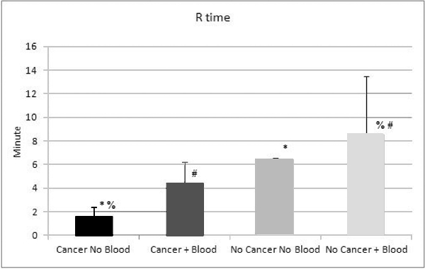
* p=0.05 % p=0.003 # p=0.02
Back to Annual Meeting Program
|

