
INVERSE EXPRESSION OF COX-1 AND COX-2 IN THE PATHOGENESIS OF BARRETT'S ADENOCARCINOMA
Publishing Number: 542
Daniel Vallboehmer, Jeffery H. Peters, Sylke Schneider, Nahid Hamoui, Kazumi Uchida, Hidekazu Kuramochi, Daisuke Shimizu, Parakrama T. Chandrasoma, Kathleen D. Danenberg, Peter Danenberg, Tom R. DeMeester, Department of Surgery, Division of Thoracic and Foregut Surgery, University of Southern California, Los Angeles, CA, Department of Biochemistry, University of Southern California, Los Angeles, CA, Department of Pathology, University of Southern California, Los Angeles, CA, Response Genetics, Inc., Los Angeles, CA, Department of Surgery, Division of Thoracic and Foregut Surgery,University of Southern California, Los Angeles, CA
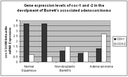
Background: Cox-2 gene expression is considered an important factor in the pathogenesis of esophageal adenocarcinoma. This conclusion is compromised by the lack of an inflammatory control in most studies and the inclusion of adjacent normal tissue. In addition cox-1, a gene thought to be constitutively expressed in most normal tissues, has not been concurrently studied. The aim of this study is to measure the expression of both genes in laser capture microdissected tissues involved in inflammation and the metaplastic-dysplastic-carcinoma sequence of Barrett's adenocarcinoma. Experimental Design: Histologic sections from endoscopic biopsies or esophagectomy specimens were classified as: reflux-esophagitis (n=8), non-dysplastic Barrett's (n=37), dysplastic Barrett's (n=17) and adenocarcinoma (n=38). Histology of the tissue of interest was confirmed by a pathologist and isolated by laser capture microdissection. Cellular RNA was extracted and reverse transcribed to cDNA. Expression levels of cox-1 and -2 and were measured by quantitative real-time PCR (Taqman®). Gene expression levels were compared to proximal esophageal squamous (n=45) epithelium of patients without Barrett's or cancer. Results: Gene expression levels of cox-2 were significantly increased in the epithelium from non-dysplastic Barrett's (p=0.003), dysplastic Barrett's (p=0.001) and adenocarcinoma (p<0.001) compared to normal squamous epithelium. In contrast expression levels of cox-1 were significantly decreased in the epithelium from non-dysplastic Barrett's (p<0.001), dysplastic Barrett's (p<0.001) and adenocarcinoma (p<0.001). Expression of neither gene was changed in reflux esophagitis. Conclusion: In microdissected tissue we have confirmed the upregulation of cox-2 and it appears to be a neoplastic rather than an inflammatory phenomenon. Cox-1 is not constitutively but variably expressed in carcinogenesis, suggesting its downregulation is simultaneous with upregulation of cox-2.
 500 Cummings Center
 +1 978-927-8330
 +1 978-524-0461
