# 101213 Abstract ID: 101213 Bilious Reflux Is Especially Noxious in a Rat Model of GERD
Colman K Byrnes, Suzanne S Hilal, Petra H Nass, Tekum Fonong, Patricia M Alli, Narasimham L Parinandi, Viswanathan Natarajan, Mark D Duncan, John W Harmon, Baltimore, 21224, MD
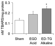
Purpose: The mechanism of esophageal injury in GERD remains poorly understood. We compared esophagitis in animals with pure duodenal reflux (ED-TG) as opposed to mixed gastro-duodenal reflux (EGD) assessing acute oxidative stress and histologic characteristics. Methods: Male 300g rats were anesthetized with intramuscular ketamine and xylazine. Mixed gastro-duodenal reflux was induced by creating a side-to-side esophago-gastro-duodenostomy (EGD), and pure duodenal reflux was produced with an end-to-end esophago-duodenostomy following a total gastrectomy (ED-TG). A third group was sham operated. Post operatively, refluxing animals received a single intraperitoneal injection of 50mg/kg iron dextran in normal saline. Animals were sacrificed at one or six weeks. The anastomosis and esophagus were examined histologically. Thiobarbituric acid reactive substances (TBARS), an index of lipid peroxidation and intracellular oxidative stress, were also quantitated on esophageal tissue lysates spectrophotometrically. Results: Following transient weight loss post-operatively, animals in all groups gained weight. The esophagus was found to be grossly thickened (4 x normal) in all duodenal reflux animals compared to both the sham operated and mixed reflux groups. On light microscopy, there was evidence of hyperkeratosis, chronic inflammation, ulceration, and papillary hyperplasia with cellular atypia extending 15 mm above the anastomosis into the mid-esophagus in the duodenal reflux group. There was evidence of increased oxidative stress in both reflux groups, but this was highest in the animals with bilious reflux (figure) *=p<0.028 ANOVA, ED-TG vs. Control. Conclusion: A purely duodenal refluxate caused more extensive and severe esophagitis than a mixed gastro-duodenal refluxate. Oxidative stress, as measured by lipid peroxidation was also greater with pure duodenal reflux.

|
 500 Cummings Center
 +1 978-927-8330
 +1 978-524-0461